MUSKELBRISTNING AF LÅRETS INDADFØRER
|
||
|
||
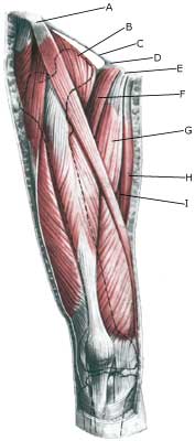
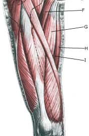
| Cause: When a muscle is subjected to a load beyond the strength of the muscle (jumping, kicking), a rupture occurs. Muscle ruptures in the groin occur most often at the fastening of the adductor longus muscle, which feels like a firm string in the groin. The rupture may be located on the fastening (where there is the greatest risk of the course being long) or in the muscular belly a few centimetres from the fastening (where experience shows that the damage heals faster). The muscle is especially damaged in sports characterised by sprinting with sudden changes of direction and sports with a lot of weight training and modest agility training (football, ice hockey), while it is rarely seen in sports characterised by agility (gymnastics). The vast majority of ruptures are partial ruptures, although total ruptures are described (article).
Symptoms: Pain upon applying pressure along the tendon with worsening upon stretching and activation of the muscle tendon (squeezing stretched legs together against resistance). In light cases a local tenderness is felt after the load (“muscle strain”, “imminent pulled muscle”). In severe cases a sudden shooting pain in the muscle is felt (partial “muscle rupture” or “pulled muscle”) and in the worst case a sudden snap is felt, rendering the muscle unusable (“total muscle rupture”). In case of total rupture a swelling can often be felt on the inside of the thigh. Acute treatment: Click here. Examination: In light cases medical examination in not necessarily required. Severe cases or cases not improved by treatment should be evaluated by a doctor so that a precise diagnosis can be made. A normal medical examination is usually sufficient in order to make the diagnosis, however, if there is any doubt concerning the diagnosis an ultrasound scan can be performed. Ultrasound is well suited to evaluate the tendons. If in the medical examination there is pain when applying pressure on the muscle attachment point in the groin, and aggravation at the same location upon stretching and activation of the adductor, there is hardly any doubt about the diagnosis and an ultrasound scan is not necessary. However, the diagnosis of groin pain can be particularly difficult (article). Treatment: Relief, stretching and slowly increasing load within the pain threshold. Complications: If progress is not smooth it should be considered whether the diagnosis is correct or whether complication have arisen. In particular the following should be considered:
Special: Shock absorbing shoes or inlays will reduce the load in the groin. In case of lack of progress or relapse after successful rehabilitation, a running style analysis can be considered to evaluate whether correction of the running style should be recommended. |