|
||
|
||
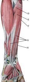
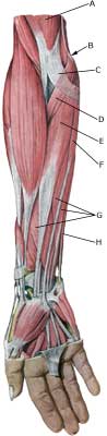
| Cause: The strength of the muscle fastening will be exceeded if subjected to repeated uniform (over)loads, causing microscopic ruptures in the tendon, and especially at the tendon fastening, resulting in an inflammation. This tendinitis is a warning that the training performed is too strenuous for the muscle tendons in question, and if the load is not reduced a chronic inflammation can arise which is problematic to treat. The condition is also called “golf elbow”, and is often a consequence of incorrect stroke technique but can be the result of a number of other causes.
Symptoms: Tenderness and pain in the area of the inner bone projection elbow (epicondylus medialis) on the elbow which is aggravated when activating the muscle group which fastens there (flexing of the wrist (flexion) against resistance and when stretching). Acute treatment: Click here. Examination: The diagnosis is usually made based on a normal medical examination, however, if there are any doubts surrounding the diagnosis an ultrasound scan can be performed which will often reveal the inflammatory changes at the muscle fastening (article). Novel use of laser doppler imaging for investigating epicondylitis. With prolonged discomfort a fraying of the bone membrane (“entesopati”) (Ultrasonic image) can be observed, as well as calcification of the soft parts which in places can have the characteristics of a calcaneal spur. Treatment: Correction of stroke technique and adjustment of equipment are naturally vital elements for a successful rehabilitation. Treatment primarily comprises relief, stretching and strength training of the forearm muscles (article). If the discomfort does not abate, the treatment can be supplemented with medicinal treatment in the form of rheumatic medicine (NSAID) or injection of corticosteroid (article). Surgical treatment can be considered if there is no change for the better, however, the results are far from convincing. Bandage: Some patients experience an improvement in the symptoms by applying tape (or a bandage) around the forearm just below the elbow (tape-instruction) Complications: If satisfactory progress is not achieved it should be considered whether the diagnosis is correct or whether complications have arisen, which can amongst others be:
|