|
||
|
||
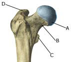
| Cause: For unknown reasons, a slow collapse of the femoral head (aseptic necrosis of the bone) can occur, resulting in a disintegration and flattening of the femoral head and consequently irritation in the hip joint. However, secondhand smoke is recognised as playing a role (article). The condition is mainly seen in children in the 3-11 age group, with boys being affected three times more often than girls. 10% of cases are on both sides of the hip.
Symptoms: There will only be slight pain initially, with tiredness in the hip joint upon movement and being subjected to load, as well as limping and constriction of movement of the joint. Constriction of movement when rotating the hip joint will often be observed. Increased pain will be felt later on, with more pronounced limping due to the shortening of the leg. The pain can occasionally be felt in the knee instead of the hip. Examination: It is important to undergo a medical examination as soon as possible so that the diagnosis can be made, as this is vital to achieve a good result from the treatment. It will often be necessary to supplement the examination with an ultrasound scan (Ultrasonic image), where bone change and fluid in the hip joint can clearly be identified (article) and possibly also x-ray examination where the late bone change can be seen (X-ray). Treatment: Treatment should be commenced as soon as ever possible, and primarily comprises intensive relief, with possible use of a wheel chair. The period of relief can last 1-2 years. An operative correction can be necessary if the disease leaves considerable damage (article). Rehabilitation of children and adolescents: The type of rehabilitation and load which can be permitted is completed dependant upon the severity of the disease. The rehabilitation should therefore be performed in close cooperation with the doctors controlling the treatment Complications: The condition can cause the risk of shortening of the leg and degenerative arthritis in the hip joint. There are, however, good chances for the articular head to heal to its normal shape, especially for the youngest children. |