Alle indlæg af urtehave_com

KONDITION

step2

Training ladder for:
CARTILAGE DAMAGE ON THE KNEE CAP
(CHONDROMALACIA PATELLAE)

STEP 2

KONDITION
Unlimited: Cycling. Swimming. Light jogging.

UDSPÆNDING
(10 min)

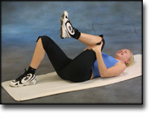
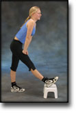
Lie on your back. Draw the injured leg up towards your head so that the muscles in the back of the thigh become increasingly stretched. Perform the exercise with outstretched as well as bent knee. Hold the position for 20 seconds and relax for 20 seconds before repeating. The exercise can also be performed standing with the injured leg outstretched on a chair while the upper body is bent slightly forwards.

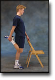
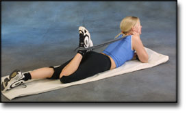
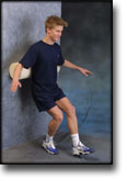
Stand with support from the back of a chair or the wall. Using your hand, bend the knee and draw the foot up and your knee slightly backwards so that the muscles in the front of the thigh become increasingly stretched. Hold the position for 20 seconds and relax for 20 seconds before repeating. The exercise can also be done lying down. If you lie on your stomach you can draw the foot up by using a towel.

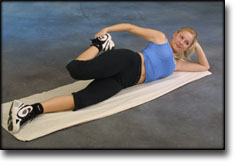
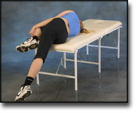
Lie on your side on a table. Bend one leg up under your body and let the other hang over the edge of the table so that the muscles in the outer side of the thigh become increasingly stretched. Hold the position for 20 seconds and relax for 20 seconds before repeating. The exercise can also be done standing by placing the outstretched injured leg behind the good leg at the same time as bending over the injured leg.

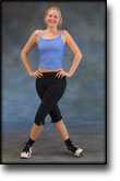
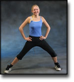
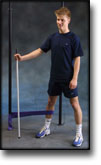
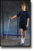
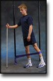
Stand with one leg outstretched and the other slightly bent. Thrust your weight to the side over the bent leg so that the inner side of the opposite thigh becomes increasingly stretched. Hold the position for 20 seconds and relax for 20 seconds before repeating.

KOORDINATION
(5 min)

Seesaw. Balance on two legs, possibly using a hand as support against the wall, balancing subsequently on one leg without support. Look straight ahead and keep knees bent.

STYRKE
(35 min)

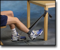
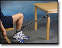
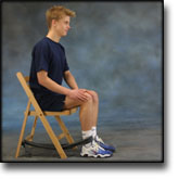
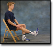
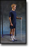
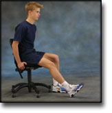
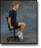
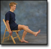
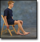
Sit on a chair with elastic around the ankle, facing the elastic. Lift the leg and slowly bend and stretch the knee.

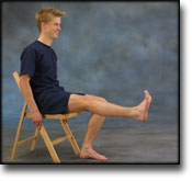
Sit on a chair with elastic attached to the ankle. Raise the leg and slowly stretch and bend the knee.

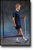
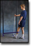
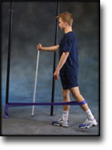
Stand with the elastic around the injured leg, facing away from the elastic. Move the leg forwards and slowly backwards. The elastic can be moved up and down the leg depending upon the strength of the knee – the stronger the knee, the lower the elastic should be.

Stand with the elastic around the injured leg, facing towards the elastic. Move the leg backwards and slowly forwards. The elastic can be moved up and down the leg depending upon the strength of the knee – the stronger the knee, the lower the elastic should be.

Stand on the healthy leg with the elastic around the inside of the injured leg. Move the injured leg from side to side in a slow smooth movement. Moving the position of the elastic lower down the leg can increase the load.

Stand on the healthy leg with the elastic around the outside of the injured leg. Move the injured leg from side to side in a slow smooth movement. Moving the position of the elastic lower down the leg can increase the load.

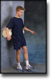
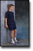
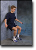
Stand with your back against a wall with a ball or firm round cushion between the wall and your back. Slowly go down to bend your knee 90 degrees before slowly rising up again.

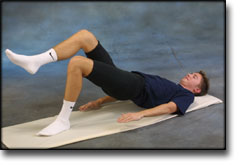
Lie on your back and bend the injured leg. Lift the healthy leg, and then raise your backside with the weight on the injured leg to be trained. Hold the position a few seconds and lower your backside again. Moving the injured leg further away from the body will increase the load.

Stand in a doorway or against a wall. Stand with your weight on the healthy leg and press the injured leg against the wall. Hold the pressure for approx. 10 seconds. Rest for 10 seconds. Repeat the exercise for approx. 3 minutes.

Stretching is carried out in the following way: stretch the muscle group for 3-5 seconds. Relax for 3-5 seconds. The muscle group should subsequently be stretched for 20 seconds. The muscle is allowed to be tender, but must not hurt. Relax for 20 seconds, after which the procedure can be repeated. The time consumed for stretching, coordination and strength training can be altered depending on the training opportunities available and individual requirements.

KONDITION

step1

Training ladder for:
CARTILAGE DAMAGE ON THE KNEE CAP
(CHONDROMALACIA PATELLAE)

STEP 1

The indications of time after stretching, coordination training and strength training show the division of time for the respective type of training when training for a period of one hour. The time indications are therefore not a definition of the daily training needs, as the daily training is determined on an individual basis.

KONDITION
Unlimited: Cycling with raised saddle. Swimming. Running in deep water.

UDSPÆNDING
(10 min)

Lie on your back. Draw the injured leg up towards your head so that the muscles in the back of the thigh become increasingly stretched. Perform the exercise with outstretched as well as bent knee. Hold the position for 20 seconds and relax for 20 seconds before repeating. The exercise can also be performed standing with the injured leg outstretched on a chair while the upper body is bent slightly forwards.

Stand with support from the back of a chair or the wall. Using your hand, bend the knee and draw the foot up and your knee slightly backwards so that the muscles in the front of the thigh become increasingly stretched. Hold the position for 20 seconds and relax for 20 seconds before repeating. The exercise can also be done lying down. If you lie on your stomach you can draw the foot up by using a towel.

Lie on your side on a table. Bend one leg up under your body and let the other hang over the edge of the table so that the muscles in the outer side of the thigh become increasingly stretched. Hold the position for 20 seconds and relax for 20 seconds before repeating. The exercise can also be done standing by placing the outstretched injured leg behind the good leg at the same time as bending over the injured leg.

Stand with one leg outstretched and the other slightly bent. Thrust your weight to the side over the bent leg so that the inner side of the opposite thigh becomes increasingly stretched. Hold the position for 20 seconds and relax for 20 seconds before repeating.

KOORDINATION
(5 min)

Seesaw. Balance on two legs, possibly using a hand as support against the wall, balancing subsequently on one leg without support. Look straight ahead and keep knees bent.

STYRKE
(35 min)

Sit on a chair with elastic around the ankle, facing the elastic. Lift the leg and slowly bend and stretch the knee.

Sit on a chair with elastic attached to the ankle. Raise the leg and slowly stretch and bend the knee.

Stand with the elastic around the injured leg, facing away from the elastic. Move the leg forwards and slowly backwards. The elastic can be moved up and down the leg depending upon the strength of the knee – the stronger the knee, the lower the elastic should be.

Stand with the elastic around the injured leg, facing towards the elastic. Move the leg backwards and slowly forwards. The elastic can be moved up and down the leg depending upon the strength of the knee – the stronger the knee, the lower the elastic should be.

Stand on the healthy leg with the elastic around the inside of the injured leg. Move the injured leg from side to side in a slow smooth movement. Moving the position of the elastic lower down the leg can increase the load.

Stand on the healthy leg with the elastic around the outside of the injured leg. Move the injured leg from side to side in a slow smooth movement. Moving the position of the elastic lower down the leg can increase the load.

Stand with your back to the wall with your weight on both feet. Slowly go down and bend the knee to 90 degrees, and slowly rise again.

Lie on your back and bend the injured leg. Lift the healthy leg, and then raise your backside with the weight on the injured leg to be trained. Hold the position a few seconds and lower your backside again. Moving the injured leg further away from the body will increase the load.

Stand in a doorway or against a wall. Stand with your weight on the healthy leg and press the injured leg against the wall. Hold the pressure for approx. 10 seconds. Rest for 10 seconds. Repeat the exercise for approx. 3 minutes.

Stretching is carried out in the following way: stretch the muscle group for 3-5 seconds. Relax for 3-5 seconds. The muscle group should subsequently be stretched for 20 seconds. The muscle is allowed to be tender, but must not hurt. Relax for 20 seconds, after which the procedure can be repeated. The time consumed for stretching, coordination and strength training can be altered depending on the training opportunities available and individual requirements.

treatment-article

SportNetDoc

“Dem bones”: osteochondral injuries of the knee.

Hinshaw MH, Tuite MJ, De Smet AA. Magn Reson Imaging Clin N Am 2000 May;8(2):335-48.

MR imaging plays a valuable role in the diagnosis and staging of osteochondral injuries of the femorotibial joint. Bone contusions may be the source of a patient’s pain, and MR imaging characteristics of certain types may help to predict which contusions might progress to more serious osteochondral lesions. MR imaging also is vital in the diagnosis of occult osteochondral fractures and in accurately classifying displaced intra-articular fractures. Although osteochondral dissecans usually is diagnosed radiographically, MR imaging is the best noninvasive test for determining if an osteochondral fragment is unstable. Unstable lesions are a treatable cause of knee pain.

KONDITION

step4

Training ladder for:
DEGENERATIVE ARTHRITIS
(ARTROSE)

STEP 4

KONDITION
Unlimited: Cycling with raised saddle. Swimming. Running and spurting with sudden directional change and jumping.

UDSPÆNDING
(10 min)

Lie on your back. Draw the injured leg up towards your head so that the muscles in the back of the thigh become increasingly stretched. Perform the exercise with outstretched as well as bent knee. Hold the position for 20 seconds and relax for 20 seconds before repeating. The exercise can also be performed standing with the injured leg outstretched on a chair while the upper body is bent slightly forwards.

Stand with support from the back of a chair or the wall. Using your hand, bend the knee and draw the foot up and your knee slightly backwards so that the muscles in the front of the thigh become increasingly stretched. Hold the position for 20 seconds and relax for 20 seconds before repeating. The exercise can also be done lying down. If you lie on your stomach you can draw the foot up by using a towel.

Lie on your side on a table. Bend one leg up under your body and let the other hang over the edge of the table so that the muscles in the outer side of the thigh become increasingly stretched. Hold the position for 20 seconds and relax for 20 seconds before repeating. The exercise can also be done standing by placing the outstretched injured leg behind the good leg at the same time as bending over the injured leg.

Stand with one leg outstretched and the other slightly bent. Thrust your weight to the side over the bent leg so that the inner side of the opposite thigh becomes increasingly stretched. Hold the position for 20 seconds and relax for 20 seconds before repeating.

KOORDINATION
(5 min)

Seesaw. Balance on two legs, possibly using a hand as support against the wall, balancing subsequently on one leg without support. Look straight ahead and keep knees bent.

STYRKE
(45 min)

Sit on a chair with elastic around the ankle, facing the elastic. Lift the leg and slowly bend and stretch the knee.

Stand on the healthy leg with the elastic around the inside of the injured leg. Move the injured leg from side to side in a slow smooth movement. Moving the position of the elastic lower down the leg can increase the load.

Stand on the healthy leg with the elastic around the outside of the injured leg. Move the injured leg from side to side in a slow smooth movement. Moving the position of the elastic lower down the leg can increase the load.

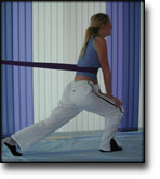
Stand with elastic around the hip. Step forward over one knee and hold the front foot firmly against the floor. Bend the rear leg and go forward onto your toes. Remember to change leg.

Stand with your back against a wall with a ball or firm round cushion between the wall and your back. Slowly go down to bend your knee 90 degrees before slowly rising up again.

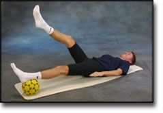
Lie on your back with a ball or firm round cushion under the injured leg. Lift your backside up from the floor and stretch the healthy leg. Hold the position for a few seconds.

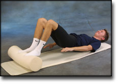
Lie on your back with a ball or firm round cushion under both feet. Roll the ball backwards and forwards in a steady pace while lifting your backside.

Stretching is carried out in the following way: stretch the muscle group for 3-5 seconds. Relax for 3-5 seconds. The muscle group should subsequently be stretched for 20 seconds. The muscle is allowed to be tender, but must not hurt. Relax for 20 seconds, after which the procedure can be repeated. The time consumed for stretching, coordination and strength training can be altered depending on the training opportunities available and individual requirements.

KONDITION

step2

Training ladder for:
DEGENERATIVE ARTHRITIS
(ARTROSE)

STEP 2

KONDITION
Unlimited: Cycling. Swimming. Light jogging.

UDSPÆNDING
(10 min)

Lie on your back. Draw the injured leg up towards your head so that the muscles in the back of the thigh become increasingly stretched. Perform the exercise with outstretched as well as bent knee. Hold the position for 20 seconds and relax for 20 seconds before repeating. The exercise can also be performed standing with the injured leg outstretched on a chair while the upper body is bent slightly forwards.

Stand with support from the back of a chair or the wall. Using your hand, bend the knee and draw the foot up and your knee slightly backwards so that the muscles in the front of the thigh become increasingly stretched. Hold the position for 20 seconds and relax for 20 seconds before repeating. The exercise can also be done lying down. If you lie on your stomach you can draw the foot up by using a towel.

Lie on your side on a table. Bend one leg up under your body and let the other hang over the edge of the table so that the muscles in the outer side of the thigh become increasingly stretched. Hold the position for 20 seconds and relax for 20 seconds before repeating. The exercise can also be done standing by placing the outstretched injured leg behind the good leg at the same time as bending over the injured leg.

Stand with one leg outstretched and the other slightly bent. Thrust your weight to the side over the bent leg so that the inner side of the opposite thigh becomes increasingly stretched. Hold the position for 20 seconds and relax for 20 seconds before repeating.

KOORDINATION
(5 min)

Seesaw. Balance on two legs, possibly using a hand as support against the wall, balancing subsequently on one leg without support. Look straight ahead and keep knees bent.

STYRKE
(45 min)

Sit on a chair with elastic around the ankle, facing the elastic. Lift the leg and slowly bend and stretch the knee.

Sit on a chair with elastic attached to the ankle. Raise the leg and slowly stretch and bend the knee.

Stand on the healthy leg with the elastic around the inside of the injured leg. Move the injured leg from side to side in a slow smooth movement. Moving the position of the elastic lower down the leg can increase the load.

Stand on the healthy leg with the elastic around the outside of the injured leg. Move the injured leg from side to side in a slow smooth movement. Moving the position of the elastic lower down the leg can increase the load.

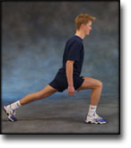
Go forward on the injured leg until the knee is bent to max. 90 degrees. Stand up on the same leg and return to the starting position.

Stand on the injured leg up on a stool with the elastic around your waist. Stand facing the wall. Walk backwards up and down from the stool under resistance from the elastic, alternating between right and left leg first.

Lie on your back with a ball or firm round cushion under both feet. Roll the ball backwards and forwards in a steady pace while lifting your backside.

Stretching is carried out in the following way: stretch the muscle group for 3-5 seconds. Relax for 3-5 seconds. The muscle group should subsequently be stretched for 20 seconds. The muscle is allowed to be tender, but must not hurt. Relax for 20 seconds, after which the procedure can be repeated. The time consumed for stretching, coordination and strength training can be altered depending on the training opportunities available and individual requirements.

KONDITION

step1

Training ladder for:
DEGENERATIVE ARTHRITIS
(ARTROSE)

STEP 1

The indications of time after stretching, coordination training and strength training show the division of time for the respective type of training when training for a period of one hour. The time indications are therefore not a definition of the daily training needs, as the daily training is determined on an individual basis.

KONDITION
Unlimited: Cycling with raised saddle. Swimming. Running in deep water.

UDSPÆNDING
(10 min)

Lie on your back. Draw the injured leg up towards your head so that the muscles in the back of the thigh become increasingly stretched. Perform the exercise with outstretched as well as bent knee. Hold the position for 20 seconds and relax for 20 seconds before repeating. The exercise can also be performed standing with the injured leg outstretched on a chair while the upper body is bent slightly forwards.

Stand with support from the back of a chair or the wall. Using your hand, bend the knee and draw the foot up and your knee slightly backwards so that the muscles in the front of the thigh become increasingly stretched. Hold the position for 20 seconds and relax for 20 seconds before repeating. The exercise can also be done lying down. If you lie on your stomach you can draw the foot up by using a towel.

Lie on your side on a table. Bend one leg up under your body and let the other hang over the edge of the table so that the muscles in the outer side of the thigh become increasingly stretched. Hold the position for 20 seconds and relax for 20 seconds before repeating. The exercise can also be done standing by placing the outstretched injured leg behind the good leg at the same time as bending over the injured leg.

Stand with one leg outstretched and the other slightly bent. Thrust your weight to the side over the bent leg so that the inner side of the opposite thigh becomes increasingly stretched. Hold the position for 20 seconds and relax for 20 seconds before repeating.

KOORDINATION
(5 min)

Seesaw. Balance on two legs, possibly using a hand as support against the wall, balancing subsequently on one leg without support. Look straight ahead and keep knees bent.

STYRKE
(45 min)

Sit on a chair with elastic around the ankle, facing the elastic. Lift the leg and slowly bend and stretch the knee.

Sit on a chair with elastic attached to the ankle. Raise the leg and slowly stretch and bend the knee.

Pull yourself forward on a chair by use of your heel against the floor. Increase the resistance by having someone hold the chair.

Sit on a chair and lift the knee to a horizontal position. Hold for 1 minute, lower the leg to approx. 45 degrees for 30 seconds. Lower again to the starting position.

Stand on the healthy leg with the elastic around the inside of the injured leg. Move the injured leg from side to side in a slow smooth movement. Moving the position of the elastic lower down the leg can increase the load.

Stand on the healthy leg with the elastic around the outside of the injured leg. Move the injured leg from side to side in a slow smooth movement. Moving the position of the elastic lower down the leg can increase the load.

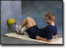
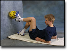
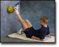
Lie on your back with the healthy leg bent and the injured leg against the wall. Place a ball between the foot and the wall. Move the injured leg up and down the wall while applying a slight pressure on the ball.

Stretching is carried out in the following way: stretch the muscle group for 3-5 seconds. Relax for 3-5 seconds. The muscle group should subsequently be stretched for 20 seconds. The muscle is allowed to be tender, but must not hurt. Relax for 20 seconds, after which the procedure can be repeated. The time consumed for stretching, coordination and strength training can be altered depending on the training opportunities available and individual requirements.

treatment-article

SportNetDoc

Effects of muscle-strength training on the functional status of patients with osteoarthritis of the knee joint.

Schilke JM, Johnson GO, Housh TJ, O’Dell JR. Nurs Res 1996 Mar-Apr;45(2):68-72.

This study was designed to determine whether an 8-week isokinetic muscle-strength-training program improved the functional health status of patients with osteoarthritis of the knee joint. Twenty volunteers with osteoarthritis of the knee joint were randomly assigned to either an experimental (n=10) or control (n=10) group. The experimental group completed six sets of five maximal contractions three times per week for 8 weeks on a Cybex II dynamometer at 90 degrees per second. Both groups were pre- and posttest for extension and flexion strength of the right and left legs, the 50-foot walk time, range of motion at the knee joint, the Osteoarthritis Screening Index (OASI), and the Arthritis Impact Measurement Scale (AIMS). There was a significant decrease in pain and stiffness, and a significant increase in mobility. There was also a significant decline in arthritis activity in the experimental group as measured by the OASI and AIMS. The experimental group significantly increased in all strength measures, while the control group increased in only right leg flexion and left leg extension across the training period.

KONDITION

step3

Training ladder for:
DEGENERATIVE ARTHRITIS
(ARTROSE)

STEP 3

KONDITION
Unlimited: Cycling with raised saddle. Swimming. Running with increasing speed and cautious directional change.

UDSPÆNDING
(10 min)

Lie on your back. Draw the injured leg up towards your head so that the muscles in the back of the thigh become increasingly stretched. Perform the exercise with outstretched as well as bent knee. Hold the position for 20 seconds and relax for 20 seconds before repeating. The exercise can also be performed standing with the injured leg outstretched on a chair while the upper body is bent slightly forwards.

Stand with support from the back of a chair or the wall. Using your hand, bend the knee and draw the foot up and your knee slightly backwards so that the muscles in the front of the thigh become increasingly stretched. Hold the position for 20 seconds and relax for 20 seconds before repeating. The exercise can also be done lying down. If you lie on your stomach you can draw the foot up by using a towel.

Lie on your side on a table. Bend one leg up under your body and let the other hang over the edge of the table so that the muscles in the outer side of the thigh become increasingly stretched. Hold the position for 20 seconds and relax for 20 seconds before repeating. The exercise can also be done standing by placing the outstretched injured leg behind the good leg at the same time as bending over the injured leg.

Stand with one leg outstretched and the other slightly bent. Thrust your weight to the side over the bent leg so that the inner side of the opposite thigh becomes increasingly stretched. Hold the position for 20 seconds and relax for 20 seconds before repeating.

KOORDINATION
(5 min)

Seesaw. Balance on two legs, possibly using a hand as support against the wall, balancing subsequently on one leg without support. Look straight ahead and keep knees bent.

STYRKE
(45 min)

Sit on a chair with elastic around the ankle, facing the elastic. Lift the leg and slowly bend and stretch the knee.

Stand on the healthy leg with the elastic around the inside of the injured leg. Move the injured leg from side to side in a slow smooth movement. Moving the position of the elastic lower down the leg can increase the load.

Stand on the healthy leg with the elastic around the outside of the injured leg. Move the injured leg from side to side in a slow smooth movement. Moving the position of the elastic lower down the leg can increase the load.

Stand with elastic around the hip. Step forward over one knee and hold the front foot firmly against the floor. Bend the rear leg and go forward onto your toes. Remember to change leg.

Stand with your back to the wall with your weight on both feet. Slowly go down and bend the knee to 90 degrees, and slowly rise again.

Lie on your back with a ball or firm round cushion under the injured leg. Lift your backside up from the floor and stretch the healthy leg. Hold the position for a few seconds.

Lie on your back with a ball or firm round cushion under both feet. Roll the ball backwards and forwards in a steady pace while lifting your backside.

Stretching is carried out in the following way: stretch the muscle group for 3-5 seconds. Relax for 3-5 seconds. The muscle group should subsequently be stretched for 20 seconds. The muscle is allowed to be tender, but must not hurt. Relax for 20 seconds, after which the procedure can be repeated. The time consumed for stretching, coordination and strength training can be altered depending on the training opportunities available and individual requirements.

article

SportNetDoc

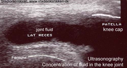
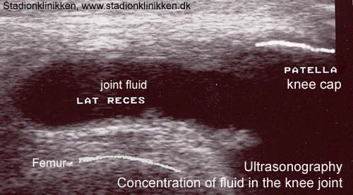
Placement of intra-articular injections verified by ultrasonography and injected air as contrast medium.

Fredberg U, van Overeem Hansen G, Bolvig L. Ann Rheum Dis 2001 May;60(5):542.

Intraarticular injection of long-acting corticosteroid is a corner stone in rheumatological treatment. The injected intra-articular corticoid is more effective when the placement is correctly secured. Injection of radiographic contrast material has shown that less than half of the injections are correct placed in the joint space after blind injection. Generally, the clinical application of ultrasonographic examinations can be enhanced by contrast agents. The most common used technique is creation of microbubble contrast agents. Such agents, applied to the bloodstream, have been used for hepatic, nephrologic, cardiologic and transcranial examinations. Obviously, the risk of air embolism depends on the anatomic site of the injected air contrast. Transient ischemic attacks are described after echocardiography with air contrast and in animal models haemodynamic effects during venous air infusion can be measured. Intra-articular injection of air and subsequently lateral and posterior radiographs has shown that this technique can enhance the procedure precision. The disadvantage of this method is that the result can first be seen after the injection, and that a correction can only be made with a new injection. In the joint space the air is separated from the vascular system and when only small amounts of sterile air are used the risk of venous air embolism is neglectable. Air is a very effective contrast medium in ultrasonography. Air sonography has been used for the diagnosis of meniscus lesions in knee joints and for rotator cuff lesions in the shoulder. We expand the applicability of this method to all joints, not only for diagnostic purposes, but also for the correct placement of the needle before injection of medicine (steroid, osmium-acid, viscosupplementation): The sterile air that is contained in the capped vial with lidocain or steroid is used as contrast medium. The needle is ultrasonographic guided placed in the joint space profound for the distended capsule. When the steroid and lidocain are mixed in the syringe a small volume of air will be in the needle itself (˜ 0.05 ml). The air in the needle is clearly seen when the injection is started and will secure the correct placement of the needle. With this technique, it is not necessary to use two separate syringes and the inclination of the syringe will not cause the air to move from the needle to the bottom of the syringe. If the knee is injected, injection directly into the recess is recommended, which will make the small volume of air possible to be seen momentary. Figure 1 illustrates the ultrasonography of a MTP joint in a patient before (UL-34A) and after (UL-34B) injected air. The intraarticular air is clearly seen. We had made more than one thousand ultrasonography guided intraarticular injection without any complications. This method is easy, inexpensive, without risk and radiation and should be used routinely in rheumatology. Especially chemical synovectomy of the knee should always be guided by ultrasonography and with this technique also smaller joins can be considered for chemical synovectomy.