Alle indlæg af urtehave_com

cause-article1

SportNetDoc

Incidence and causes of tenosynovitis of the wrist extensors in long distance paddle canoeists.

du Toit P, Sole G, Bowerbank P, Noakes TD. Br J Sports Med 1999 Apr;33(2):105-9.

OBJECTIVES.
To investigate the incidence and causes of acute tenosynovitis of the forearm of long distance canoeists.

METHOD.
A systematic sample of canoeists competing in four canoe marathons were interviewed. The interview included questions about the presence and severity of pain in the forearm and average training distances. Features of the paddles and canoes were determined.

RESULTS.
An average of 23% of the competitors in each race developed this condition. The incidence was significantly higher in the dominant than the nondominant hand but was unrelated to the type of canoe and the angle of the paddle blades. Canoeists who covered more than 100 km a week for eight weeks preceding the race had a significantly lower incidence of tenosynovitis than those who trained less. Environmental conditions during racing, including fast flowing water, high winds, and choppy waters, and the paddling techniques, especially hyperextension of the wrist during the pushing phase of the stroke, were both related to the incidence of tenosynovitis.

CONCLUSION.
Tenosynovitis is a common injury in long distance canoeists. The study suggests that development of tenosynovitis is not related to the equipment used, but is probably caused by difficult paddling conditions, in particular uneven surface conditions, which may cause an altered paddling style. However, a number of factors can affect canoeing style. Level of fitness and the ability to balance even a less stable canoe, thereby maintaining optimum paddling style without repeated eccentric loading of the forearm tendons to limit hyperextension of the wrist, would seem to be important

examination-article

SportNetDoc

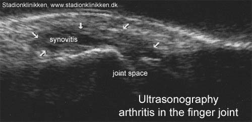
Usefulness of high resolution US in the evaluation of effusion in osteoarthritic first carpometacarpal joint.

Iagnocco A, Coari G. Scand J Rheumatol 2000;29(3):170-3.

OBJECTIVE.
The aim of this study is to provide a reproducible and quantitative sonographic method for evaluation of effusion in the first carpometacarpal joint in osteoarthritis.

METHODS.
High resolution sonography of the carpometacarpal joint of the thumb was carried out in 20 normal joints and in 57 joints from patients with osteoarthritis. A 10 MHz transducer was used.

RESULTS.
The articular cavity appeared as a hypoechoic triangular area. In normal joints the mean values obtained by measuring the distance between the apex and the base of the triangle was 2.89 mm (SD 0.22). In osteoarthritic joints it was significantly increased (p<0.001). The authors assert that the presence of effusion is very likely if the value is >3.33 mm (mean+2 SD).

CONCLUSIONS.
Sonography provides useful, reproducible, and quantitative data for detection of effusion within the first carpometacarpal joint.

tape-instruction

Tapening


Type: THUMB BANDAGE:

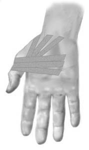
Objective: Stabilise the metacarpophalangeal joint (MCP1 joint)

Application: The thumb should be held in a neutral position. Start in the middle of the palm adjacent to the wrist, draw the tape around the thumb’s metacarpophalangeal joint and return to the palm and cross the start tape (A). The next tape is applied in similar fashion, but started slightly further out on the palm (B). A third strip can be applied in a continued fan formation. (C).

treatment-article2

SportNetDoc

Role of MR imaging in the management of “skier’s thumb” injuries.

Plancher KD, Ho CP, Cofield SS, Viola R, Hawkins RJ. Magn Reson Imaging Clin N Am 1999 Feb;7(1):73-84, viii.

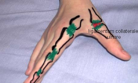
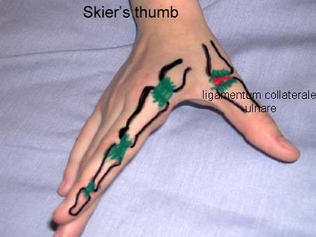
“Skier’s thumb” is an acute rupture of the ulnar collateral ligament (UCL) of the metacarpophalangeal (MCP) joint of the thumb. As the method of choice in evaluating soft tissue injuries, MR imaging is useful in evaluating UCL injuries. This article reviews current concepts regarding the rupture of the UCL, including a study of 34 UCL injuries in which MR imaging was used as the main diagnostic tool. When correlated with surgical findings, MR imaging resulted in identifying UCL tears with 96% sensitivity and 95% specificity.

treatment-article1

SportNetDoc

Sonography of the injured ulnar collateral ligament of the thumb.

Hergan K, Mittler C. J Bone Joint Surg Br 1995 Jan;77(1):77-83.

We used high-resolution ultrasonography to image the ulnar collateral ligament in 39 patients who had sustained recent injuries of the metacarpophalangeal joint of the thumb. All the patients were subsequently operated on and the lesions of the ligament were recorded. In 36 patients the preoperative ultrasonographic diagnosis was correct. Five of these showed no rupture of the ligament. In the other 31, ultrasonography correctly distinguished between rupture in situ (15) and rupture with dislocation of the ligament (16). Misdiagnosis by ultrasonography in three cases was due to delay of the investigation (three weeks after injury) in one, to technical error in one and to misinterpretation of the image in one.

examination-article

SportNetDoc

The skier’s thumb.

Heim D. Acta Orthop Belg 1999 Dec;65(4):440-6.

The incidence of skier’s thumb (rupture of the ulnar collateral ligament of the first metacarpophalangeal joint) is increasing. To determine whether conservative or surgical treatment is indicated, ultrasound (US) and magnetic resonance imaging (MRI) have been advocated in the last few years. Surgery should be performed in the case of an unstable joint with a ligamentous tear or in the presence of a displaced bony fragment. Several techniques for surgical repair in acute and old ruptures are proposed. Conservative and postoperative treatment consists of immobilization of the joint in a splint or thumb spica cast for 4 weeks. The best results are obtained in bony avulsion fractures. Conservative treatment of lesions requiring surgical treatment may result in permanent disability of the joint; thus, correct diagnosis is mandatory.

KONDITION

step4

Training ladder for:
INFLAMMATION OF THE HOLLOW FOOT TENDON
(FASCIITIS PLANTARIS)

STEP 4

KONDITION
Unlimited: Cycling. Swimming.

UDSPÆNDING
(10 min)

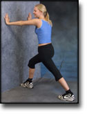
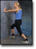
Stand with the injured leg stretched backwards with the toes facing front. Slowly bend the knee so that the calf muscles become increasingly stretched. Hold the position for 20 seconds and relax for 20 seconds before repeating.

Stand with the injured leg outstretched with the ankle joint bent up against the wall. Press your abdomen against the wall so that the calf muscles become increasingly stretched. Hold the position for 20 seconds and relax for 20 seconds before repeating.

KOORDINATION
(5 min)

Seesaw. Balance on two legs, possibly using a hand as support against the wall, balancing subsequently on one leg without support. Look straight ahead and keep knees bent.

STYRKE
(45 min)

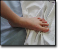
Sit with your bare toes on a tea towel and curl the tea towel together using your toes.

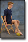
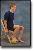
Sit with a tennis ball under the foot. Roll the ball backwards and forwards and from side to side.

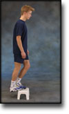
Stand with both forefeet on a stool or doorstep with the heel out over the edge. Slowly rise up on to your toes with your weight on the healthy leg. Go slowly down on the injured leg as far as you can go. Use the healthy leg to rise up on to your toes again. The exercise should be performed with stretched, as well as bent knee. Wearing a rucksack and gradually increasing the ballast in the rucksack can increase the load.

Stand on a soft surface. Rise slowly up on tiptoe and go down again.

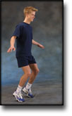
Stand on your toes with bent knees. Place your weight forward on the toes and keep your balance.

Stretching is carried out in the following way: stretch the muscle group for 3-5 seconds. Relax for 3-5 seconds. The muscle group should subsequently be stretched for 20 seconds. The muscle is allowed to be tender, but must not hurt. Relax for 20 seconds, after which the procedure can be repeated. The time consumed for stretching, coordination and strength training can be altered depending on the training opportunities available and individual requirements.

KONDITION

step3

Training ladder for:
INFLAMMATION OF THE HOLLOW FOOT TENDON
(FASCIITIS PLANTARIS)

STEP 3

KONDITION
Unlimited: Cycling. Swimming.

UDSPÆNDING
(10 min)

Stand with the injured leg stretched backwards with the toes facing front. Slowly bend the knee so that the calf muscles become increasingly stretched. Hold the position for 20 seconds and relax for 20 seconds before repeating.

Stand with the injured leg outstretched with the ankle joint bent up against the wall. Press your abdomen against the wall so that the calf muscles become increasingly stretched. Hold the position for 20 seconds and relax for 20 seconds before repeating.

KOORDINATION
(5 min)

Seesaw. Balance on two legs, possibly using a hand as support against the wall, balancing subsequently on one leg without support. Look straight ahead and keep knees bent.

STYRKE
(45 min)

Sit with your bare toes on a tea towel and curl the tea towel together using your toes.

Sit with a tennis ball under the foot. Roll the ball backwards and forwards and from side to side.

Stand with both forefeet on a stool or doorstep with the heel out over the edge. Slowly rise up on to your toes with your weight on the healthy leg. Go slowly down on the injured leg as far as you can go. Use the healthy leg to rise up on to your toes again. The exercise should be performed with stretched, as well as bent knee. Wearing a rucksack and gradually increasing the ballast in the rucksack can increase the load.

Stand on a soft surface. Rise slowly up on tiptoe and go down again.

Stand on your toes with bent knees. Place your weight forward on the toes and keep your balance.

Stretching is carried out in the following way: stretch the muscle group for 3-5 seconds. Relax for 3-5 seconds. The muscle group should subsequently be stretched for 20 seconds. The muscle is allowed to be tender, but must not hurt. Relax for 20 seconds, after which the procedure can be repeated. The time consumed for stretching, coordination and strength training can be altered depending on the training opportunities available and individual requirements.