Alle indlæg af urtehave_com

The Diagnosis


A precise diagnosis can be made in far the majority of cases from the case history and a clinical examination. In many cases it will, however, be advantageous to supplement with:

X-ray, which is primarily used for suspicions of fractures and bone membrane tears (avulsion). The examination entails exposure to x-ray irradiation.

CT-scanning, which is used with certain fractures as a supplement to ordinary x-ray examination. The examination entails marked exposure to x-ray irradiation.

MRI-scanning, which is primarily indicated in cases where further examination of the conditions inside the joints is desired – since ultrasound scanning is easier, considerably cheaper and presumed just as effective in evaluating conditions outside of the joints. The examination entails exposure to magnetic irradiation, which is, as far as anyone knows, harmless.

Diagnostic blockade with local anaesthetic has only modest risks associated with it if performed with the correct injection technique (possibly under guidance of ultrasound), and can contribute with considerable diagnostic information.

Arthroscopy, which is used for diagnostic examination of the conditions in the joint if it is not possible to determine “from outside” what is wrong in the joint despite use of all other modern technology. This method is associated with a number of risks in relation to the other examination methods.

Ultrasound scanning, which is the cheapest, quickest and most suitable examination to enhance the findings from a clinical examination. The examination entails exposure to ultrasound irradiation which is completely harmless.

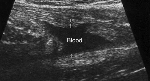
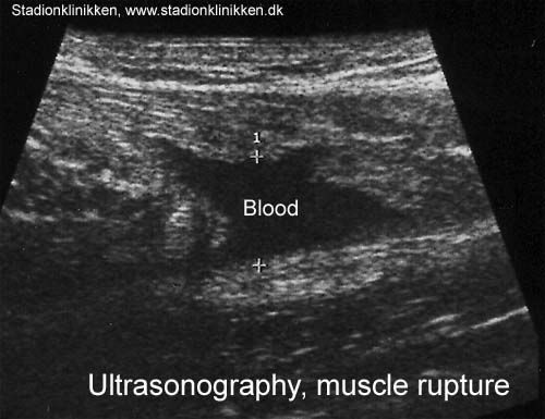
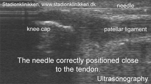
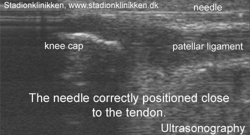
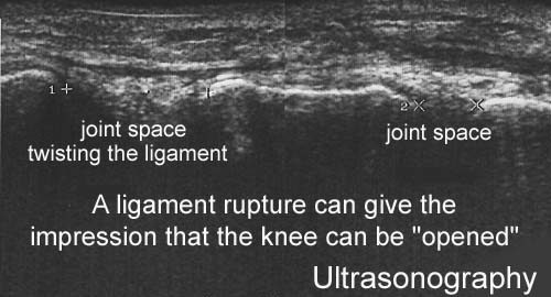
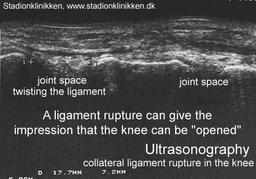
Ultrasound scanning has developed rapidly over the recent decades. Ultrasound scanning is today an integral part of the clinical examination associated with sports medicine and other rheumatology related conditions, and acts as the physician’s “extended hand”. Ultrasound scanning will generally contribute to clarifying matters where the diagnosis is not certain, or the course of events is not as could be expected. Ultrasound scanning is particularly suitable for revealing the structures outside, and within the joints. The examination allows evaluation of the muscles, tendons and joints when activated (dynamic ultrasound scanning) (Ultrasonic image) as well as ultrasound-guided diagnostic and therapeutic draining of accumulations (blood, joint fluid, etc.). Furthermore, ultrasound-guided injection provides the possibility of placing the needle within a range of precision of 1/10 mm (Ultrasonic image). Injections which have earlier been performed “blind” have only been successfully placed at the desired location in under half of the attempts, and in more than 50% of the cases have erroneously hit other structures. It is for this reason that all injections where error in placement involves certain risks (injection around the large tendons and in the vicinity of large vessels and nerves) should always be performed under guidance of ultrasound. The correct placement of the needle can be performed by direct visualization of the tip of the needle in the most instances, but a small volume of sterile air (less than one drop) acts as an effective contrast medium and can ascertain a 100% correct placement of the needle in all injections, (article), (Ultrasonic image).

Examples where ultrasound scanning has enabled precise diagnosis and treatment:

Case history 1: Stress fracture (Ultrasonic image)
Case history 2: Meniscus lesion, (Ultrasonic image)

Case history 3: Bursitis of the tendon of the knee (Baker’s cyst) (Ultrasonic image)

Case history 4: Injection, Jumper’s knee (Ultrasonic image), (Ultrasonic image), (Ultrasonic image).


Case history 5: Myositis ossificans, (Ultrasonic image)

KONDITION

step4

Training ladder for:
MUSCLE RUPTURE IN THE CALF
(RUPTURA MUSCULI)

STEP 4

KONDITION
Unlimited: Cycling. Swimming. Running and spurting with jumps.

UDSPÆNDING
(10 min)

Stand with the injured leg stretched backwards with the toes facing front. Slowly bend the knee so that the calf muscles become increasingly stretched. Hold the position for 20 seconds and relax for 20 seconds before repeating.

Stand with the injured leg outstretched with the ankle joint bent up against the wall. Press your abdomen against the wall so that the calf muscles become increasingly stretched. Hold the position for 20 seconds and relax for 20 seconds before repeating.

Stand on the good leg while gaining support on the wall with the hand on the same side. Bend the knee of the injured leg and draw the heel towards the buttocks. Take hold of the big toe side of the foot and draw slowly upwards so that the ankle joint is stretched to the maximum and drawn slightly outwards so that increased stretching is experienced on the inside of the shin bone. Hold the position for 20 seconds and relax for 20 seconds before repeating.

Stand on the good leg while gaining support from the wall with the opposite hand. Bend the knee of the injured leg and draw the heel towards the buttocks. Take hold of the little toe side of the foot and draw slowly upwards so that the ankle joint is stretched to the maximum and drawn over the good leg so that increased stretching is experienced on the outer side of the shin bone. Hold the position for 20 seconds and relax for 20 seconds before repeating.

KOORDINATION
(5 min)

Stand on the leg to be trained. Take-off and land on the same leg.

Seesaw. Balance on two legs, possibly using a hand as support against the wall, balancing subsequently on one leg without support. Look straight ahead and keep knees bent.

STYRKE
(45 min)

Sit on the floor. Tip the foot up and down with elastic under the forefoot so that the elastic becomes taut.

Sit on the floor. Tip the foot up and down with elastic on top of the forefoot so that the elastic becomes taut.

Sit on the floor. Tip the foot from side to side with elastic on the outer side of the foot, without moving the knee.

Sit on the floor. Tip the foot from side to side with elastic on the inner side of the foot, without moving the knee.

Stand on a soft surface. Rise slowly up on tiptoe and go down again.

Stand behind a chair. Rise slowly up on tiptoe and go down again.

Go up and down from the stool. Go up with alternating right and left legs.

Stand on the healthy leg with elastic fixed around the hip. The elastic should be fixed to the wall or a wall bar. Take-off on the healthy leg and land on the leg to be trained and keep your balance. Remember that the elastic should be positioned so that it gives resistance at the moment of take-off. Change legs.

Stand with both legs on the stool with elastic around the hip. Take-off and land with feet together.

Stand with feet together. Using the ankle joint to take off, hop approx. 5 cm and land on both feet. The exercise should be done on one leg when you are able to do it without discomfort using both legs.

Stretching is carried out in the following way: stretch the muscle group for 3-5 seconds. Relax for 3-5 seconds. The muscle group should subsequently be stretched for 20 seconds. The muscle is allowed to be tender, but must not hurt. Relax for 20 seconds, after which the procedure can be repeated. The time consumed for stretching, coordination and strength training can be altered depending on the training opportunities available and individual requirements.

KONDITION

step3

Training ladder for:
MUSCLE RUPTURE IN THE CALF
(RUPTURA MUSCULI)

STEP 3

KONDITION
Unlimited: Cycling. Swimming. Running with increasing speed and skipping.

UDSPÆNDING
(10 min)

Stand with the injured leg stretched backwards with the toes facing front. Slowly bend the knee so that the calf muscles become increasingly stretched. Hold the position for 20 seconds and relax for 20 seconds before repeating.

Stand with the injured leg outstretched with the ankle joint bent up against the wall. Press your abdomen against the wall so that the calf muscles become increasingly stretched. Hold the position for 20 seconds and relax for 20 seconds before repeating.

Stand on the good leg while gaining support on the wall with the hand on the same side. Bend the knee of the injured leg and draw the heel towards the buttocks. Take hold of the big toe side of the foot and draw slowly upwards so that the ankle joint is stretched to the maximum and drawn slightly outwards so that increased stretching is experienced on the inside of the shin bone. Hold the position for 20 seconds and relax for 20 seconds before repeating.

Stand on the good leg while gaining support from the wall with the opposite hand. Bend the knee of the injured leg and draw the heel towards the buttocks. Take hold of the little toe side of the foot and draw slowly upwards so that the ankle joint is stretched to the maximum and drawn over the good leg so that increased stretching is experienced on the outer side of the shin bone. Hold the position for 20 seconds and relax for 20 seconds before repeating.

KOORDINATION
(5 min)

Seesaw. Balance on two legs, possibly using a hand as support against the wall, balancing subsequently on one leg without support. Look straight ahead and keep knees bent.

STYRKE
(45 min)

Sit on the floor. Tip the foot up and down with elastic under the forefoot so that the elastic becomes taut.

Sit on the floor. Tip the foot up and down with elastic on top of the forefoot so that the elastic becomes taut.

Sit on the floor. Tip the foot from side to side with elastic on the outer side of the foot, without moving the knee.

Sit on the floor. Tip the foot from side to side with elastic on the inner side of the foot, without moving the knee.

Stand on a soft surface. Rise slowly up on tiptoe and go down again.

Stand behind a chair. Rise slowly up on tiptoe and go down again.

Go up and down from the stool. Go up with alternating right and left legs.

Stand on the healthy leg with elastic fixed around the hip. The elastic should be fixed to the wall or a wall bar. Take-off on the healthy leg and land on the leg to be trained and keep your balance. Remember that the elastic should be positioned so that it gives resistance at the moment of take-off. Change legs.

Stand with both legs on the stool with elastic around the hip. Take-off and land with feet together.

Stretching is carried out in the following way: stretch the muscle group for 3-5 seconds. Relax for 3-5 seconds. The muscle group should subsequently be stretched for 20 seconds. The muscle is allowed to be tender, but must not hurt. Relax for 20 seconds, after which the procedure can be repeated. The time consumed for stretching, coordination and strength training can be altered depending on the training opportunities available and individual requirements.

KONDITION

step2

Training ladder for:
MUSCLE RUPTURE IN THE CALF
(RUPTURA MUSCULI)

STEP 2

KONDITION
Unlimited: Cycling. Swimming. Light jogging.

UDSPÆNDING
(10 min)

Stand with the injured leg stretched backwards with the toes facing front. Slowly bend the knee so that the calf muscles become increasingly stretched. Hold the position for 20 seconds and relax for 20 seconds before repeating.

Stand with the injured leg outstretched with the ankle joint bent up against the wall. Press your abdomen against the wall so that the calf muscles become increasingly stretched. Hold the position for 20 seconds and relax for 20 seconds before repeating.

Stand on the good leg while gaining support on the wall with the hand on the same side. Bend the knee of the injured leg and draw the heel towards the buttocks. Take hold of the big toe side of the foot and draw slowly upwards so that the ankle joint is stretched to the maximum and drawn slightly outwards so that increased stretching is experienced on the inside of the shin bone. Hold the position for 20 seconds and relax for 20 seconds before repeating.

Stand on the good leg while gaining support from the wall with the opposite hand. Bend the knee of the injured leg and draw the heel towards the buttocks. Take hold of the little toe side of the foot and draw slowly upwards so that the ankle joint is stretched to the maximum and drawn over the good leg so that increased stretching is experienced on the outer side of the shin bone. Hold the position for 20 seconds and relax for 20 seconds before repeating.

KOORDINATION
(5 min)

Seesaw. Balance on two legs, possibly using a hand as support against the wall, balancing subsequently on one leg without support. Look straight ahead and keep knees bent.

STYRKE
(45 min)

Sit on the floor. Tip the foot up and down with elastic under the forefoot so that the elastic becomes taut.

Sit on the floor. Tip the foot up and down with elastic on top of the forefoot so that the elastic becomes taut.

Sit on the floor. Tip the foot from side to side with elastic on the outer side of the foot, without moving the knee.

Sit on the floor. Tip the foot from side to side with elastic on the inner side of the foot, without moving the knee.

Stand on a soft surface. Rise slowly up on tiptoe and go down again.

Stand behind a chair. Rise slowly up on tiptoe and go down again.

Stretching is carried out in the following way: stretch the muscle group for 3-5 seconds. Relax for 3-5 seconds. The muscle group should subsequently be stretched for 20 seconds. The muscle is allowed to be tender, but must not hurt. Relax for 20 seconds, after which the procedure can be repeated. The time consumed for stretching, coordination and strength training can be altered depending on the training opportunities available and individual requirements.

KONDITION

step1

Training ladder for:
MUSCLE RUPTURE IN THE CALF
(RUPTURA MUSCULI)

STEP 1

The indications of time after stretching, coordination training and strength training show the division of time for the respective type of training when training for a period of one hour. The time indications are therefore not a definition of the daily training needs, as the daily training is determined on an individual basis.

KONDITION
Unlimited: Cycling. Swimming. Running in deep water.

UDSPÆNDING
(10 min)

Stand with the injured leg stretched backwards with the toes facing front. Slowly bend the knee so that the calf muscles become increasingly stretched. Hold the position for 20 seconds and relax for 20 seconds before repeating.

Stand with the injured leg outstretched with the ankle joint bent up against the wall. Press your abdomen against the wall so that the calf muscles become increasingly stretched. Hold the position for 20 seconds and relax for 20 seconds before repeating.

Stand on the good leg while gaining support on the wall with the hand on the same side. Bend the knee of the injured leg and draw the heel towards the buttocks. Take hold of the big toe side of the foot and draw slowly upwards so that the ankle joint is stretched to the maximum and drawn slightly outwards so that increased stretching is experienced on the inside of the shin bone. Hold the position for 20 seconds and relax for 20 seconds before repeating.

Stand on the good leg while gaining support from the wall with the opposite hand. Bend the knee of the injured leg and draw the heel towards the buttocks. Take hold of the little toe side of the foot and draw slowly upwards so that the ankle joint is stretched to the maximum and drawn over the good leg so that increased stretching is experienced on the outer side of the shin bone. Hold the position for 20 seconds and relax for 20 seconds before repeating.

KOORDINATION
(5 min)

Seesaw. Balance on two legs, possibly using a hand as support against the wall, balancing subsequently on one leg without support. Look straight ahead and keep knees bent.

STYRKE
(45 min)

Sit on the floor. Tip the foot up and down with elastic under the forefoot so that the elastic becomes taut.

Sit on the floor. Tip the foot from side to side with elastic on the outer side of the foot, without moving the knee.

Sit on the floor. Tip the foot from side to side with elastic on the inner side of the foot, without moving the knee.

Lay on the floor with a cushion under the calf. Tip the foot up and down without putting any resistance on the foot.

Lay on your back with your foot above heart-height. Tip the foot up and down and from side to side.

Sit on a chair. Keep the heel firmly on the ground and tip the toes up.

Stretching is carried out in the following way: stretch the muscle group for 3-5 seconds. Relax for 3-5 seconds. The muscle group should subsequently be stretched for 20 seconds. The muscle is allowed to be tender, but must not hurt. Relax for 20 seconds, after which the procedure can be repeated. The time consumed for stretching, coordination and strength training can be altered depending on the training opportunities available and individual requirements.

treatment-article

SportNetDoc

Acute tears of the medial head of the gastrocnemius.

Shields CL Jr, Redix L, Brewster CE. Foot Ankle 1985 Jan-Feb;5(4):186-90.

Twenty-five patients with acute tears of the medial head of the gastrocnemius were evaluated in follow-up from 1 to 3 years after injury. The patients were treated with a heel lift, calf sleeve, and physical therapy. The majority of the patients were men who injured their dominant lower extremity in one explosive episode. Cybex II testing revealed no significant difference in the plantarflexion strength of the noninjured and injured extremity after healing. All patients returned to their previous level of athletic activity.