Alle indlæg af urtehave_com

KONDITION

STEP3

GENOPTRÆNING

TRAINING LADDER FOR CHILDREN AND ADOLESCENTS:
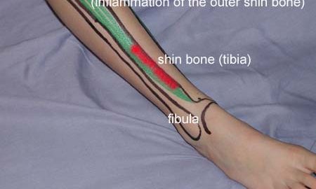
FOR INFLAMMATION OF THE OUTER SHIN BONE
(LATERAL TIBIAL PERIOSTITIS)

STEP 3

The following rehabilitation program will cover the needs of the vast majority of children with inflammation of the outer shinbone. Older teenagers involved in sports at a high level can advantageously use the rehabilitation program for adults.
KONDITION
Unlimited: Cycling. Swimming. Running with increasing speed and skipping.

UDSPÆNDING
(10 min)

Stand on the good leg while gaining support on the wall with the hand on the same side. Bend the knee of the injured leg and draw the heel towards the buttocks. Take hold of the big toe side of the foot and draw slowly upwards so that the ankle joint is stretched to the maximum and drawn slightly outwards so that increased stretching is experienced on the inside of the shin bone. Hold the position for 20 seconds and relax for 20 seconds before repeating.

Stand on the good leg while gaining support from the wall with the opposite hand. Bend the knee of the injured leg and draw the heel towards the buttocks. Take hold of the little toe side of the foot and draw slowly upwards so that the ankle joint is stretched to the maximum and drawn over the good leg so that increased stretching is experienced on the outer side of the shin bone. Hold the position for 20 seconds and relax for 20 seconds before repeating.

KOORDINATION
(5 min)

Seesaw. Balance on two legs, possibly using a hand as support against the wall, balancing subsequently on one leg without support. Look straight ahead and keep knees bent.

STYRKE
(15 min)

Sit on the floor. Tip the foot up and down with elastic under the forefoot so that the elastic becomes taut.

Sit on the floor. Tip the foot up and down with elastic on top of the forefoot so that the elastic becomes taut.

Sit on the floor. Tip the foot from side to side with elastic on the outer side of the foot, without moving the knee.

Sit on the floor. Tip the foot from side to side with elastic on the inner side of the foot, without moving the knee.

Stretching is carried out in the following way: stretch the muscle group for 3-5 seconds. Relax for 3-5 seconds. The muscle group should subsequently be stretched for 20 seconds. The muscle is allowed to be tender, but must not hurt. Relax for 20 seconds, after which the procedure can be repeated.

The time consumed for stretching, coordination and strength training can be altered depending on the training opportunities available and individual requirements.

KONDITION

STEP2

GENOPTRÆNING

TRAINING LADDER FOR CHILDREN AND ADOLESCENTS:
FOR INFLAMMATION OF THE OUTER SHIN BONE
(LATERAL TIBIAL PERIOSTITIS)

STEP 2

The following rehabilitation program will cover the needs of the vast majority of children with inflammation of the outer shinbone. Older teenagers involved in sports at a high level can advantageously use the rehabilitation program for adults.
KONDITION
Unlimited: Cycling. Swimming. Light jogging.

UDSPÆNDING
(5 min)

Stand on the good leg while gaining support from the wall with the opposite hand. Bend the knee of the injured leg and draw the heel towards the buttocks. Take hold of the little toe side of the foot and draw slowly upwards so that the ankle joint is stretched to the maximum and drawn over the good leg so that increased stretching is experienced on the outer side of the shin bone. Hold the position for 20 seconds and relax for 20 seconds before repeating.

STYRKE
(15 min)

Sit on the floor. Tip the foot up and down with elastic under the forefoot so that the elastic becomes taut.

Sit on the floor. Tip the foot up and down with elastic on top of the forefoot so that the elastic becomes taut.

Sit on the floor. Tip the foot from side to side with elastic on the outer side of the foot, without moving the knee.

Sit on the floor. Tip the foot from side to side with elastic on the inner side of the foot, without moving the knee.

Stretching is carried out in the following way: stretch the muscle group for 3-5 seconds. Relax for 3-5 seconds. The muscle group should subsequently be stretched for 20 seconds. The muscle is allowed to be tender, but must not hurt. Relax for 20 seconds, after which the procedure can be repeated.

The time consumed for stretching, coordination and strength training can be altered depending on the training opportunities available and individual requirements.

KONDITION

STEP1

GENOPTRÆNING

TRAINING LADDER FOR CHILDREN AND ADOLESCENTS:
FOR INFLAMMATION OF THE OUTER SHIN BONE
(LATERAL TIBIAL PERIOSTITIS)

STEP 1

The following rehabilitation program will cover the needs of the vast majority of children with inflammation of the outer shinbone. Older teenagers involved in sports at a high level can advantageously use the rehabilitation program for adults.
KONDITION
Unlimited: Cycling. Swimming. Running in deep water.

UDSPÆNDING
(5 min)

Stand on the good leg while gaining support from the wall with the opposite hand. Bend the knee of the injured leg and draw the heel towards the buttocks. Take hold of the little toe side of the foot and draw slowly upwards so that the ankle joint is stretched to the maximum and drawn over the good leg so that increased stretching is experienced on the outer side of the shin bone. Hold the position for 20 seconds and relax for 20 seconds before repeating.

STYRKE
(5 min)

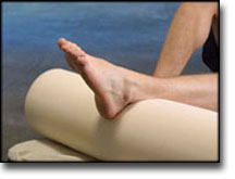
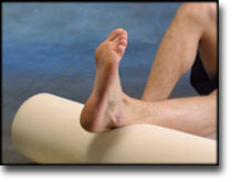
Lay on the floor with a cushion under the calf. Tip the foot up and down without putting any resistance on the foot.

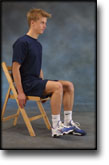
Sit on a chair. Keep the heel firmly on the ground and tip the toes up.

Stretching is carried out in the following way: stretch the muscle group for 3-5 seconds. Relax for 3-5 seconds. The muscle group should subsequently be stretched for 20 seconds. The muscle is allowed to be tender, but must not hurt. Relax for 20 seconds, after which the procedure can be repeated.

The time consumed for stretching, coordination and strength training can be altered depending on the training opportunities available and individual requirements.

complications-article

SportNetDoc

Skiing injuries in children: lower leg fractures.

Ungerholm S, Gierup J, Lindsjo U, Magnusson A. Int J Sports Med 1985 Oct;6(5):292-7

A study was made of 113 children who sustained a lower leg fracture during downhill skiing. The age distribution showed a peak between 4 and 7 years in both sexes, and boys had another peak in their teens. Three of four patients were beginners. To a large extent, they had had their bindings adjusted in ski shops. Three of four bindings did not release at the time of the accident. Spiral fractures in the shaft or distal metaphysis of the tibia predominated (73%); the incidence of concomitant fibular fracture was low. The degree of malalignment was generally small. The results of treatment, which was generally conservative, were good, 7% of the patients having minor sequelae 1-3 years after the accident. To reduce the risks in children’s downhill skiing, the following measures seem important: intensified training during the beginner stage; increased supervision by parents and in ski schools; development of children’s release bindings and testing methods; and adequate instruction of personnel in ski shops.

treatment-article

SportNetDoc

Intramedullary Kirschner wiring for tibia fractures in children.

Qidwai SA. J Pediatr Orthop 2001 May-Jun;21(3):294-7

This is a retrospective analysis of the results of 84 tibial fractures in children treated by intramedullary Kirschner wiring. Thirty were open fractures (9 grade I, 10 grade II, 8 grade IIIA, 3 grade IIIB). There were 65 boys and 18 girls with an average age of 10.23 years (range 4-15). The patient was placed supine on an orthopedic traction table. Under fluoroscopic control, two Kirschner wires (2.5-3.5-mm thick) were introduced antegrade from proximal metaphysis (level of tibial tuberosity) to distal metaphysis, one each from medial and lateral cortices. Open fractures were stabilized after meticulous wound debridement. Average time to union was 9.5 weeks (range 8-14). None developed delayed union. However, one grade IIIB open fracture progressed to infected nonunion; it healed after an autogenous bone graft. No infections were seen in closed fractures, but four superficial and one deep infection occurred in open fractures. Closed intramedullary Kirschner wire fixation for unstable or open tibial fractures in children is a simple surgical technique that produces good clinical and functional results.

cause-article

SportNetDoc

Isolated fractures of the tibia with intact fibula in children: a review of 95 patients.

Yang JP, Letts RM. J Pediatr Orthop 1997 May-Jun;17(3):347-51

Isolated tibial fractures with an intact fibula are the most common tibial fracture pattern in children. When displaced, this fracture can be difficult to reduce and retain in the position because of the splinting of the intact fibula. A 4-year review of 95 children with fracture of the tibia with an intact fibula was performed at the Children’s Hospital of Eastern Ontario. Eighty-one percent of the fractures were caused by an indirect rotational twisting force. Seventy-three percent of fractures were localized at the distal third of the tibial shaft. Varus angulation deformity occurred most commonly when the fracture line started distally on the anteromedial side of the tibia and progressed in an oblique or spiral manner to the proximal posterolateral aspect of the tibia. This was postulated to be caused by the posterior flexor muscle forces being more concentrated medially, whereas laterally, the intact fibula acts like a splint, thus producing a bending moment resulting in varus angulation. Close follow-up and monitoring of the isolated tibial fractures with weekly radiographs for the first 3 weeks is recommended.

rehabilitation-article

SportNetDoc

Femoral shaft stress fractures in athletes.

Hershman EB, Lombardo J, Bergfeld JA. Clin Sports Med 1990 Jan;9(1):111-9.

Stress fractures of the femoral shaft in athletes occur most commonly in the proximal third of the femur. They can, however, also be found in the mid- or distal third. Conservative treatment is highly successful in healing these fractures without complications. Athletes can usually return to activity in 8 to 14 weeks. Recognition of the symptoms characteristic of these fractures (vague thigh pain, diffuse tenderness, no trauma) will assist early diagnosis. Early definitive diagnosis can be made by radionuclide scanning or later, by plain radiography, if symptoms have been present for a sufficient period. Diagnosis is not limited to novice runners since runners with significant mileage, or baseball or basketball players, can develop femoral shaft stress fractures.

treatment-article

SportNetDoc

Interventions for preventing and treating stress fractures and stress reactions of bone of the lower limbs in young adults.

Gillespie WJ, Grant I. Cochrane Database Syst Rev 2000;(2):CD000450.

BACKGROUND.
Stress reaction in bone, which may proceed to a fracture, is a significant problem in military recruits and in athletes, particularly long distance runners.

OBJECTIVES.
To evaluate the evidence from controlled trials of treatments and programmes for prevention or management of lower limb stress fractures and stress reactions of bone in active young adults.

SEARCH STRATEGY.
We searched the Cochrane Musculoskeletal Injuries Group Trials Register, The Cochrane Library, MEDLINE, EMBASE, Current Contents, Dissertation Abstracts, Index to UK Theses and the bibliographies of identified articles. Date of last search: December 1997.

SELECTION CRITERIA.
Any randomised or quasi-randomised trial evaluating a programme or treatment to prevent or treat lower limb stress reactions of bone or stress fractures in active young adults.

DATA COLLECTION AND ANALYSIS.
Searching, a decision on inclusion or exclusion, methodological assessment, and data extraction were carried out according to a predetermined protocol included in the body of the review. Analysis using Review Manager software allowed pooling of data and calculation of Peto odds ratios and absolute risk reductions, each with 95% confidence intervals.

MAIN RESULTS.
The use of “shock absorbing” insoles, evaluated in four trials, appears to reduce the incidence of stress fractures and stress reactions of bone (Peto odds ratio 0.47, 95% confidence interval 0. 30 to 0.76). Incomplete data from one trial indicated that reduction of running and jumping intensity may also be effective. The use of pneumatic braces in the rehabilitation of tibial stress fractures significantly reduces the time to recommencing training (weighted mean difference -42.6 days, 95% confidence interval -55.8 to -29.4 days).

REVIEWER’S CONCLUSIONS.
The use of shock absorbing insoles in footwear reduces the incidence of stress fractures in athletes and military personnel. Rehabilitation after tibial stress fracture is aided by the use of pneumatic bracing.

examination-article2

SportNetDoc

Femoral stress fractures.

Boden BP, Speer KP. Clin Sports Med 1997 Apr;16(2):307-17.

Stress fractures are common overuse injuries attributed to the repetitive trauma associated with vigorous weightbearing activities. A high index of suspicion is necessary to diagnose stress fractures of the femur because the symptoms may be vague. The precipitating factors, whether related to training errors or medical conditions, should be thoroughly evaluated. Early diagnosis of distraction femoral neck stress fractures is critical to avoid serious complications. Femoral shaft stress fractures have excellent healing potential when diagnosed early and treated non-operatively. Stress fractures of the femoral condyles are uncommon, but should be included in the differential of knee pain.