Kategoriarkiv: Foot

Bone fracture in the forefoot

Diagnosis: BONE FRACTURE IN THE FOREFOOT


Anatomy:
The foot bones comprise the 7 tarsal bones (ossa tarsi), the 5 metatarsal bones (ossa metatarsi) and the 14 bones in the toes (phalanx).

  1. Phalanx media
  2. Tuberositas ossis metatarsalis V
  3. Os cuboideum
  4. Calcaneus
  5. Talus
  6. Os naviculare
  7. Os cuneiforme laterale
  8. Os cuneiforme intermedium
  9. Os cuneiforme mediale
  10. Os metatarsalei
  11. Os sesamoideum
  12. Phalanx proximalis
  13. Phalanx distalis

THE FOOT FROM ABOVE

Cause: A blow or violent twist can cause a fracture of the bone (X-ray picture).

Symptoms: Pain when applying pressure (direct or indirect tenderness), and when applying load or strain.

Acute treatment: Click here.

Examination: X-ray examination will usually reveal the fracture. The fracture can in some cases first be seen after 14 days, thus the x-ray examination should be repeated if there is a continued suspicion of a fracture.

Treatment: Treatment is completely dependent upon which bones are broken, and whether there is a dislocation of the fracture. In some cases relief and rest without bandaging can be opted for, whereas other types of fracture require bandaging and possibly surgical intervention (article).

Rehabilitation: Rehabilitation is totally dependent upon the type of fracture, and the treatment (conservative or surgical). Also read rehabilitation, general

Complications: If there is not a steady improvement in the condition a medical examination should be performed once more to ensure that the fracture is healing according to plan. In some cases, a false joint can develop which will require (renewed) surgical treatment (X-ray picture), (X-ray picture).

Special: As there is a risk that the injury can cause permanent disability, all cases should be reported to your insurance company.

KONDITION

STEP4

TRAINING LADDER FOR CHILDREN AND ADOLESCENTS:
FOR INFLAMMATION OF THE HOLLOW FOOT TENDON
(FASCIITIS PLANTARIS)

STEP 4

The majority of young patients can manage with relief until the pain subsides, following which the sports activity can be slowly resumed. The following rehabilitation program will cover the needs for the vast majority of children with inflammation of the hollow foot tendon. Older teenagers involved in sports at a high level can advantageously use the rehabilitation program for adults.
KONDITION
Unlimited: Cycling. Swimming. Running with increasing distance on a soft surface.

UDSPÆNDING
(5 min)

Stand with the injured leg stretched backwards with the toes facing front. Slowly bend the knee so that the calf muscles become increasingly stretched. Hold the position for 20 seconds and relax for 20 seconds before repeating.

Stand with the injured leg outstretched with the ankle joint bent up against the wall. Press your abdomen against the wall so that the calf muscles become increasingly stretched. Hold the position for 20 seconds and relax for 20 seconds before repeating.

KOORDINATION
(5 min)

Seesaw. Balance on two legs, possibly using a hand as support against the wall, balancing subsequently on one leg without support. Look straight ahead and keep knees bent.

STYRKE
(10 min)

Sit with your bare toes on a tea towel and curl the tea towel together using your toes.

Stand with both forefeet on a stool or doorstep with the heel out over the edge. Slowly rise up on to your toes with your weight on the healthy leg. Go slowly down on the injured leg as far as you can go. Use the healthy leg to rise up on to your toes again. The exercise should be performed with stretched, as well as bent knee. Wearing a rucksack and gradually increasing the ballast in the rucksack can increase the load.

Stand on your toes with bent knees. Place your weight forward on the toes and keep your balance.

Stretching is carried out in the following way: stretch the muscle group for 3-5 seconds. Relax for 3-5 seconds. The muscle group should subsequently be stretched for 20 seconds. The muscle is allowed to be tender, but must not hurt. Relax for 20 seconds, after which the procedure can be repeated.

The time consumed for stretching, coordination and strength training can be altered depending on the training opportunities available and individual requirements.

KONDITION

STEP3

TRAINING LADDER FOR CHILDREN AND ADOLESCENTS:
FOR INFLAMMATION OF THE HOLLOW FOOT TENDON
(FASCIITIS PLANTARIS)

STEP 3

The majority of young patients can manage with relief until the pain subsides, following which the sports activity can be slowly resumed. The following rehabilitation program will cover the needs for the vast majority of children with inflammation of the hollow foot tendon. Older teenagers involved in sports at a high level can advantageously use the rehabilitation program for adults.
KONDITION
Unlimited: Cycling. Swimming. Light jogging (few minutes) on a soft surface.

UDSPÆNDING
(5 min)

Stand with the injured leg stretched backwards with the toes facing front. Slowly bend the knee so that the calf muscles become increasingly stretched. Hold the position for 20 seconds and relax for 20 seconds before repeating.

Stand with the injured leg outstretched with the ankle joint bent up against the wall. Press your abdomen against the wall so that the calf muscles become increasingly stretched. Hold the position for 20 seconds and relax for 20 seconds before repeating.

STYRKE
(45 min)

Sit with your bare toes on a tea towel and curl the tea towel together using your toes.

Stand with both forefeet on a stool or doorstep with the heel out over the edge. Slowly rise up on to your toes with your weight on the healthy leg. Go slowly down on the injured leg as far as you can go. Use the healthy leg to rise up on to your toes again. The exercise should be performed with stretched, as well as bent knee. Wearing a rucksack and gradually increasing the ballast in the rucksack can increase the load.

Stretching is carried out in the following way: stretch the muscle group for 3-5 seconds. Relax for 3-5 seconds. The muscle group should subsequently be stretched for 20 seconds. The muscle is allowed to be tender, but must not hurt. Relax for 20 seconds, after which the procedure can be repeated.

The time consumed for stretching, coordination and strength training can be altered depending on the training opportunities available and individual requirements.

KONDITION

STEP2

TRAINING LADDER FOR CHILDREN AND ADOLESCENTS:
FOR INFLAMMATION OF THE HOLLOW FOOT TENDON
(FASCIITIS PLANTARIS)

STEP 2

The majority of young patients can manage with relief until the pain subsides, following which the sports activity can be slowly resumed. The following rehabilitation program will cover the needs for the vast majority of children with inflammation of the hollow foot tendon. Older teenagers involved in sports at a high level can advantageously use the rehabilitation program for adults.
KONDITION
Unlimited: Cycling. Swimming.

UDSPÆNDING
(5 min)

Stand with the injured leg stretched backwards with the toes facing front. Slowly bend the knee so that the calf muscles become increasingly stretched. Hold the position for 20 seconds and relax for 20 seconds before repeating.

Stand with the injured leg outstretched with the ankle joint bent up against the wall. Press your abdomen against the wall so that the calf muscles become increasingly stretched. Hold the position for 20 seconds and relax for 20 seconds before repeating.

STYRKE
(5 min)

Sit with your bare toes on a tea towel and curl the tea towel together using your toes.

Stand with both forefeet on a stool or doorstep with the heel out over the edge. Slowly rise up on to your toes with your weight on the healthy leg. Go slowly down on the injured leg as far as you can go. Use the healthy leg to rise up on to your toes again. The exercise should be performed with stretched, as well as bent knee. Wearing a rucksack and gradually increasing the ballast in the rucksack can increase the load.

Stretching is carried out in the following way: stretch the muscle group for 3-5 seconds. Relax for 3-5 seconds. The muscle group should subsequently be stretched for 20 seconds. The muscle is allowed to be tender, but must not hurt. Relax for 20 seconds, after which the procedure can be repeated.

The time consumed for stretching, coordination and strength training can be altered depending on the training opportunities available and individual requirements.

KONDITION

STEP1

TRAINING LADDER FOR CHILDREN AND ADOLESCENTS:
FOR INFLAMMATION OF THE HOLLOW FOOT TENDON
(FASCIITIS PLANTARIS)

STEP 1

The majority of young patients can manage with relief until the pain subsides, following which the sports activity can be slowly resumed. The following rehabilitation program will cover the needs for the vast majority of children with inflammation of the hollow foot tendon. Older teenagers involved in sports at a high level can advantageously use the rehabilitation program for adults.
KONDITION
Unlimited: Cycling. Swimming.

UDSPÆNDING
(5 min)

Stand with the injured leg stretched backwards with the toes facing front. Slowly bend the knee so that the calf muscles become increasingly stretched. Hold the position for 20 seconds and relax for 20 seconds before repeating.

Stand with the injured leg outstretched with the ankle joint bent up against the wall. Press your abdomen against the wall so that the calf muscles become increasingly stretched. Hold the position for 20 seconds and relax for 20 seconds before repeating.

STYRKE
(5 min)

Stand with both forefeet on a stool or doorstep with the heel out over the edge. Slowly rise up on to your toes with your weight on the healthy leg. Go slowly down on the injured leg as far as you can go. Use the healthy leg to rise up on to your toes again. The exercise should be performed with stretched, as well as bent knee. Wearing a rucksack and gradually increasing the ballast in the rucksack can increase the load.

Stretching is carried out in the following way: stretch the muscle group for 3-5 seconds. Relax for 3-5 seconds. The muscle group should subsequently be stretched for 20 seconds. The muscle is allowed to be tender, but must not hurt. Relax for 20 seconds, after which the procedure can be repeated.

The time consumed for stretching, coordination and strength training can be altered depending on the training opportunities available and individual requirements.

Bandage(tape-instruction)


Type: HOLLOW FOOT TENDON:

Objective: Support, and therefore relieve, the aponeurosis under the foot (aponeurosis plantaris).

Application: A tape “anchor” is applied around the forefoot behind the toe pad (A). The second strip (B) begins from the anchor on the outer border of the foot, taken around the heel (under the heel tendon) and forward under the instep on the inside to the anchor. The third strip (C) starts from the anchor on the inner border of the foot, is taken around the heel in the same way and forward under the instep on the outer side, to the anchor. A possible fourth strip begins from the anchor on the outer border of the foot in between the two other strips, and is taken around the heel and forward under the instep between the two other strips. Finish with a further strip on top of the anchor A.

Rupture of the hollow foot tendon

Diagnosis: RUPTURE OF THE HOLLOW FOOT TENDON
(Ruptura aponeurosis plantaris)


Anatomy:
The calf muscle (M Gastrocnemicus) is comprised of two muscle heads which gather in a wide tendinous ligament and continue in to the Achilles tendon. Another of the larger calf muscles (M Soleus) is attached to the front side of the Achilles tendon and thus forms a part of the Achilles tendon. The calf muscles are attached to the rear of the heel bone (calcaneus) via the Achilles tendon. The hollow foot tendon (aponeurosis plantaris) runs from the heel bone under the sole, and is attached to all five toes. The hollow foot tendon is instrumental in maintaining the arch running the length of the foot (Photo).

  1. Aponeurosis plantaris
  2. Tuber calcanei

SOLE OF THE FOOT

Cause: Rupture of the hollow foot tendon occurs after sudden, forceful overload (for example landing after jumping). In a number of cases, a rupture of the hollow foot tendon is preceded by inflammation of the tendon (fasciitis plantaris). Since the hollow foot tendon is a functional extension of the Achilles tendon, the provoking factors which are instrumental in injuries due to overload of the hollow foot tendon and the Achilles tendon are often the same.

Symptoms: In light cases, a localised tenderness can be felt at the point of attachment of the hollow foot tendon under the heel bone, or under the arch of the foot after strain or load (“sprain”, “threatening muscle pull”). In more severe cases, sudden shooting pains can be felt in the tendon (“partial rupture”, “pulled muscle”) and at the worst a sensation of feeling and hearing a “crack” after which it is impossible to run and pain is felt whilst walking (“total tendon rupture”), where it is often possible to feel a defect in the hollow foot tendon. Total ruptures are very rare.

Acute treatment: Click here.

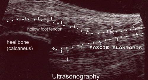
Examination: In all cases when there is a sense of a “crack”, or sudden shooting pains in the tendon, medical attention should be sought as soon as possible to make the diagnosis. Ultrasound scanning (or MRI examination) is used to advantage for making a swift and correct diagnosis, as injuries requiring treatment will often be overlooked under normal clinical examination (article).

Treatment: The chosen treatment will usually be relief until the pain disappears (article), and surgical intervention if the rehabilitation does not proceed satisfactorily (article). If a tender lump of cicatricial tissue develops after several months of rehabilitation (inflamed granuloma), treatment can be supplemented in medicinal form by rheumatic medicine (NSAID) or injection of corticosteroid in the area surrounding the inflamed lump of cicatricial tissue. Ultrasound guided injection increases the effect of the injection, as well as reduces the risks involved (article). As the injection of corticosteroid is always an element in the long term rehabilitation of a very serious, chronic injury, it is vital that the rehabilitation period lasts over several months in order to reduce the risk of a relapse or (further) rupture. The tendon is naturally unable to accommodate maximum strain or load after a prolonged injury period after only a short rehabilitation period. If the diagnosis is made by use of ultrasound scanning, the injections are performed under guidance of ultrasound, and the rehabilitation is progressed in accordance with the guidelines mentioned, then the treatment involving corticosteroid injections has very few risks connected (article-1) (article-2). It is not unusual for a rehabilitation period of six months before maximum strain or load in the form of jumping is permitted. It is vital for safety that injections are performed under guidance of ultrasound when treating chronic hollow foot tendon injuries.

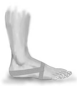
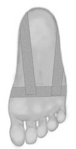
Bandage: Taping to relieve problems with the hollow foot tendon is of questionable significance, but can be attempted as the tape will not invoke further injury if applied in the correct manner (tape-instruction).

Prevention: As inflammation of the hollow foot tendon is extremely difficult to treat, it is important to prevent the injury from arising or recurring. The principles in rehabilitation, general should be followed to ensure that quickly increasing training loads at the season start, or after an injury period, are avoided. It is important that running shoes fit well (tight heel cap, shock absorbing soles). It is vital that the first signs of tenderness or pain are reacted upon, to enable the training to be adapted before the injury reaches the stage where continuing the sports activity may be at risk.

Complications: If there is not a steady improvement in the condition an ultrasound scan should be performed to exclude:

In the severe, chronic cases, all treatment and rehabilitation attempts will often result in permanent inability to continue the sports activity.

Special: As there is a risk that the injury can cause permanent disability, all cases should be reported to your insurance company.

Treatment(2)

SportNetDoc

Local corticosteroid injection in sport: review of literature and guidelines for treatment.

Fredberg U. Scand J Med Sci Sports 1997 Jun;7(3):131-9.

The risks and benefits of local injection therapy of overuse sports injuries with corticosteroids are reviewed here. Injection of corticosteroid inside the tendon has a deleterious effect on the tendon tissue and should be unanimously condemned. No reliable proof exists of the deleterious effects of peritendinous injections. Too many conclusions in the literature are based on poor scientific evidence and it is just the reiteration of a dogma if all steroid injections are abandoned. The corticosteroids represent an adjuvant treatment in the overall management of sports injuries: basic treatment is ‘active’ rest and graduated rehabilitation within the limits of pain. With proper indications there are only few and trivial complications that may occur with corticosteroid injections. Guidelines for proper local injection therapy with corticosteroids are given.

Treatment(1)

SportNetDoc

Plantar Fasciitis: Diagnosis and Conservative Management.

Gill LH. J Am Acad Orthop Surg 1997 Mar;5(2):109-117.

Plantar fasciitis is a common cause of heel pain, which frustrates patients and practitioners alike because of its resistance to treatment. It has been associated with obesity, middle age, and biomechanical abnormalities in the foot, such as tight Achilles tendon, pes cavus, and pes planus. It is considered to be most often the result of a degenerative process at the origin of the plantar fascia at the calcaneus. However, neurogenic and other causes of subcalcaneal pain are frequently cited. A combination of causative factors may be present, or the true cause may remain obscure. Although normally managed with conservative treatment, plantar fasciitis is frequently resistant to the wide variety of treatments commonly used, such as nonsteroidal anti-inflammatory drugs, rest, pads, cups, splints, orthotics, corticosteroid injections, casts, physical therapy, ice, and heat. Although there is no consensus on the efficacy of any particular conservative treatment regimen, there is agreement that nonsurgical treatment is ultimately effective in approximately 90% of patients. Since the natural history of plantar fasciitis has not been established, it is unclear how much of symptom resolution is in fact due to the wide variety of commonly used treatments.