Kategoriarkiv: Hip

clave-article2

Untitled Document

US of the painful hip in childhood: diagnostic value of cartilage thickening and muscle atrophy in the detection of Perthes disease.

Robben SG, Meradji M, Diepstraten AF, Hop WC. Radiology 1998 Jul;208(1):35-42

PURPOSE: To evaluate the combined use of several ultrasonographic (US) criteria in the detection of Perthes disease. MATERIALS AND METHODS: In a prospective study, 144 consecutive children with a painful hip underwent US. The thicknesses of the (a) anterior recess of the joint capsule, (b) cartilage of the femoral head, and (c) quadriceps muscle were assessed. Sixty-eight children with no symptoms, the control group, were also examined. RESULTS: The final diagnosis was transient synovitis (n = 58), Perthes disease (n = 21), slipped capital femoral epiphysis (SCFE; n = 5), or miscellaneous (n = 6). Fifty-four patients had no US or radiographic abnormalities, and symptoms disappeared during follow-up. The anterior recess in patients with transient synovitis was significantly wider than that in the other patients and control subjects (P < .001). Patients with Perthes disease showed significant cartilage thickening in the symptomatic hip compared with the other patients and control subjects (P < .001). Patients with Perthes disease and patients with SCFE showed significant atrophy of the ipsilateral quadriceps muscle compared with all other groups (P < .001). The combined use of these US criteria for the diagnosis of Perthes disease resulted in a positive predictive value of 95%, a negative predictive value of 95%, a sensitivity of 71%, and a specificity of 99%. CONCLUSION: The combination of several US criteria increases the diagnostic value of US of the painful hip in patients with Perthes disease.

clave-article1

Untitled Document

Secondhand smoke, hypofibrinolysis, and Legg-Perthes disease.

Glueck CJ, Freiberg RA, Crawford A, Gruppo R, Roy D, Tracy T, Sieve-Smith L, Wang P. Clin Orthop 1998 Jul;(352):159-67

In 39 children with Legg-Perthes disease who were nonsmokers, the specific aim was to assess relationships among parental cigarette smoking during pregnancy, household smoking before diagnosis of Legg-Perthes disease, hypofibrinolysis, and thrombophilia. Fifteen (38%) children had no secondhand smoke exposure; 24 (62%) had secondhand smoke exposure before their diagnosis. Seventeen (71%) of these 24 children were exposed while in utero to smoking by a parent or live in relative and also had exposure to household smoke during childhood; seven (29%) had only household smoke exposure in childhood. In the full cohort of 39 children, secondhand smoke exposure correlated inversely with the major stimulator of fibrinolysis, stimulated tissue

x-ray-artical2

Untitled Document

Osteonecrosis of the femoral head associated with slipped capital femoral epiphysis.

Kennedy JG, Hresko MT, Kasser JR, Shrock KB, Zurakowski D, Waters PM, Millis MB. J Pediatr Orthop 2001 Mar-Apr;21(2):189-93

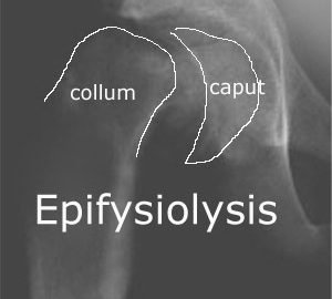
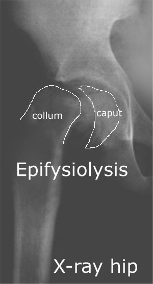
We performed a retrospective analysis of 212 patients (299 hips) with slipped capital femoral epiphysis (SCFE) over a 9-year period to assess the incidence of osteonecrosis of the femoral head. Risk factors for the occurrence of osteonecrosis and the influence of treatment on the development of osteonecrosis were determined. Osteonecrosis occurred in 4 hips with unstable SCFE (4/27) and did not occur in hips with stable SCFE (0/272). The proportion of hips in which osteonecrosis developed was significantly higher among the unstable hips (4/27 vs. 0/272, p < 0.0001). Among those with an unstable hip, younger age at presentation was a predictor of a poorer outcome. Magnitude of the slip, magnitude of reduction, and chronicity of the slip were not predictive of a poorer outcome in the unstable group. In situ fixation of the minimally or moderately displaced “unstable” SCFE demonstrated a favorable outcome. We have identified the hip at risk as an unstable SCFE. The classification of hips as unstable if the epiphysis is displaced from the metaphysis or if the patient is unable to walk is most useful in predicting a hip at risk for osteonecrosis.

x-ray-artical1

Untitled Document

Slipped capital femoral epiphysis: evaluation of different modes of treatment.

Rostoucher P, Bensahel H, Pennecot GF, Kaewpornsawan K, Mazda K. J Pediatr Orthop B 1996 Spring;5(2):96-101

We reviewed 91 hips with slipped capital femoral epiphysis (SCFE) after an average follow-up period of 6 years 6 months. Different treatment methods used, according to types and stages of slipping, are discussed. In situ fixation appears to be the best procedure for SCFE with < 60% displacement. Careful reduction-fixation is indicated in acute and acute-on-chronic SCFE > 60%. Primary rotation osteotomies are associated with a high percentage of complications. Secondary osteotomies should be simple (preferably a subtrochanteric derotation osteotomy) to reduce the risk of necrosis. Preventive contralateral fixation is indicated when the growth cartilage is still open.

x-ray-artical

Untitled Document

Diagnostic imaging of the early slipped capital femoral epiphysis.

Magnano GM, Lucigrai G, De Filippi C, Castriota Scanderberg A, Pacciani E, Toma P. Radiol Med (Torino) 1998 Jan-Feb;95(1-2):16-20

INTRODUCTION: Early slipped capital femoral epiphysis (ESCFE) can be treated surgically, with excellent results, if it is diagnosed in its initial stage; however, the rate of late or missed diagnoses remains surprisingly high. PURPOSE: We compared radiography, US and MR sensitivity in ESCFE diagnosis. MATERIAL AND METHODS: We examined 21 symptomatic overweight patients (15 boys and 6 girls) aged 9 to 15 years with anteroposterior radiographs; frog leg images were not acquired in 3 cases only. US was performed in 19 cases and the images acquired with 5-7.5 MHz probes on the sagittal plane parallel to the femoral neck. MRI was performed in 9 cases, with coronal and sagittal T1 SE and T2* GE images. RESULTS: Our sensitivity rates were 66% for anteroposterior radiography (6 false negatives), 80% for combined anteroposterior and frog leg images (3 false negatives), 95% for US (1 false negative) and 88% for MRI (1 false negative). DISCUSSION AND CONCLUSION: We believe that US is the method of choice in ESCFE diagnosis; if it is negative, but pain persists, MRI should be performed.

treatment-artical

Untitled Document

Surgery for groin and lower abdominal pain in soccer players.

Bahar A, Soudry M. Harefuah 2000 Jul;139(1-2):29-32, 78.

We treated 57 professional soccer players with groin and lower abdominal pain, 44 of them successfully, using conservative methods and 13 by surgery. Because of its anatomical site, the gracilis muscle is involved in almost all movements of the femur. It therefore is frequently involved in injury due to overuse, especially at its insertion (enthesopathy). In all 13 operated on, the gracilis was cut percutaneously, sometimes as a single procedure and sometimes with concomitant sportsmen’s inguinal hernioplasty. All except 1 of those operated on returned to their professional sport activities.

examination-article3

Untitled Document

Groin pain associated with ultrasound finding of inguinal canal posterior wall deficiency in Australian Rules footballers.

Orchard JW, Read JW, Neophyton J, Garlick D. Br J Sports Med 1998 Jun;32(2):134-9.

OBJECTIVES.
To investigate the prevalence of inguinal canal posterior wall deficiency (sports hernia) in professional Australian Rules footballers using an ultrasound technique and correlate the results with the clinical symptom of groin pain.

METHODS.
Thirty five professional Australian footballers with and without groin pain were investigated blind with a dynamic high resolution ultrasound technique for presence of posterior wall deficiency.

RESULTS.
Fourteen players had a history of significant recent groin pain and ten of these were found to have bilateral inguinal canal posterior wall deficiency (p < 0.01). The relative risk for a history of groin pain with bilateral deficiency was 8.0 (95% confidence interval 1.73 to 37.1). Groin pain was also found to be associated with increasing age (p < 0.01) which was an independent risk factor. Surgical, clinical, and ultrasound follow up for players who underwent hernia repair confirmed the validity of ultrasound as a diagnostic tool.

CONCLUSIONS.
Dynamic ultrasound examination is able to detect inguinal canal posterior wall deficiency in young males with no clinical signs of hernia. This condition is very prevalent in professional Australian Rules footballers, including some who are asymptomatic. There was a correlation between bilateral deficiency and groin pain, although the temporal relationship between the clinical and ultrasound findings is not established by the current study. Ultrasound shows promise as a diagnostic tool in athletes with chronic groin pain who are considered possible candidates for hernia repair.

examination-article2

Untitled Document

Detection of groin hernia with physical examination, ultrasound, and MRI compared with laparoscopic findings.

van den Berg JC, de Valois JC, Go PM, Rosenbusch G. Invest Radiol 1999 Dec;34(12):739-43.

OBJECTIVE.
To determine the diagnostic accuracy of physical examination, ultrasound, and dynamic MRI in patients with inguinal hernia.

METHODS.
In 41 patients with clinically evident herniations, 82 groins were evaluated using a standard ultrasound and MRI protocol, the latter including T1- and T2-weighted sequences as well as two dynamic sequences. All ultrasound examinations and MRI scans were reviewed without knowledge of clinical findings. In all cases, correlation with findings at laparoscopic surgery was made.

RESULTS.
At surgery, 55 inguinal herniations were found. Physical examination revealed 42 herniations (one false-positive finding), whereas ultrasound made the diagnosis of a hernia in 56 cases (five false-positive and four false-negative findings). MRI diagnosed 53 herniations (one false-positive and three false-negative findings). Thus, sensitivity and specificity figures were 74.5% and 96.3% for physical examination, 92.7% and 81.5% for ultrasound, and 94.5% and 96.3% for MRI.

CONCLUSIONS.
In patients with clinically uncertain herniations, MRI is a valid diagnostic tool with a high positive predictive value.

examination-article1

Untitled Document

Longstanding groin pain in athletes. A multidisciplinary approach.

Ekberg O, Persson NH, Abrahamsson PA, Westlin NE, Lilja B. Sports Med 1988 Jul;6(1):56-61.

In 21 male athletes (age 20 to 40 years) with longstanding unexplained groin pain, a multidisciplinary investigation was performed in order to reveal the underlying cause. These examinations included general surgery for detection of inguinal hernia and neuralgia, orthopaedic surgery for detection of adductor tenoperiostitis and symphysitis, urology for detection of prostatitis, radiology for performing herniography and plain film of the pelvic bones, nuclear medicine for isotope studies of the pubic bone and symphysis. In 19 patients there was a positive diagnosis for 2 or more of the diseases (10 patients had 2 diseases, 6 patients had 3 diseases, 3 patients had 4 diseases). Two patients had only signs of symphysitis. Our results show the complexity of longstanding groin pain in athletes. It also explains why therapy for one specific disease entity may fail. We conclude that this clinical setting demands the recruitment of a team with experience of different aspects of groin pain.