Kategoriarkiv: Knee

treatment-article3

SportNetDoc

Anterior cruciate ligament injuries in the skeletally immature patient.

Lo IK, Bell DM, Fowler PJ. Instr Course Lect 1998;47:351-9

Anterior cruciate ligament injury in the skeletally immature is becoming increasingly recognized and reported. History taking and physical examination based on the principles of ACL injuries in adults, with adjuncts such as arthroscopy and MRI, are effective in diagnosing ACL injury in the young patient. Evaluation of the young patient’s true level of skeletal immaturity by comparison with family growth history, examination for signs of sexual maturity, and radiographic evaluation is critical. The risk of physeal damage with surgical treatment is related to the immaturity of the distal femoral and proximal tibial physes. The functional results of nonsurgical treatment of ACL injury, either as an attempt at definitive treatment or as a temporizing plan until skeletal maturity occurs, are poor and the risks of reinjury and further meniscal and cartilage damage are significant. Surgical treatment for primary repair or extra-articular reconstruction alone has not proven to be efficacious. In the adolescent patient who is approaching skeletal maturity, risk of physeal injury is low and intra-articular reconstruction can be performed as in the adult patient. Results with respect to decreased laxity and return to athletic activities mirror those described in adults. In patients with significant growth remaining, however, surgical treatment carries much higher risks of physeal damage and subsequent deformity. Yet, as noted above, intra-articular reconstruction in truly skeletally immature patients using a soft-tissue graft through a transphyseal tibial tunnel of moderate or small diameter and the over-the-top position on the femur has not been shown to cause early physeal closure, limb-length discrepancy, or angular deformity. In humans, the maximum diameter of graft tunnel that will not cause physeal closure has not been determined Animal studies have shown that the tibial physis can be very sensitive to drilling. Therefore it is wise to use moderate tunnel diameters. Bone-patellar tendon-bone grafts have been used with success in patients closer to skeletal maturity. Their use has not been reported in the very skeletally immature knee and cannot be recommended because of the presumed high risk of physeal closure with a bone plug traversing the physis. It is hoped that improved understanding of the ACL injury in the skeletally immature patient will provide treatment options that will restore enduring knee function and prevent early arthrosis.

treatment-article2

SportNetDoc

Anterior cruciate injuries in the skeletally immature athlete: a review of treatment outcomes.

Fehnel DJ, Johnson R. Sports Med 2000 Jan;29(1):51-63

The documentation of anterior cruciate ligament (ACL) injuries in the skeletally immature athlete has significantly increased over the past decade, primarily due to increased awareness of these injuries within this younger athletic population. The evaluation of these injuries are similar to that in the adult population. Diagnostic studies such as plain radiographs, as well as magnetic resonance imaging, can delineate the location of the ACL failure. Physical presentation most commonly includes an acute haemarthrosis and ligamentous insufficiency. Several studies have demonstrated that the diagnostic reliability of the physical examination is poor in children, especially in patients less than 12 years old. The site of ACL failure in this adolescent population is most commonly at the tibial insertion. We recommend arthroscopic or arthroscopically assisted open reduction and internal fixation with nonabsorbable sutures for all displaced tibial eminence fractures. Mid-substance ACL failures also occur in this athletic age group. The association of meniscal injuries with these ACL failures appears to be greater than 50%. Historically, poor subjective and objective outcomes have been associated with primary and extra-articular repairs. Intra-articular reconstruction is the gold standard. The issue of placing the graft across open physeal plates is under investigation. Recent animal studies as well as human clinical series have demonstrated safety in placing soft tissue, i.e. hamstring grafts, across open growth plates without subsequent angular or leg length discrepancy. Historically, non-operatively treated ACL failures are associated with poor functional outcomes as well as a high incidence of meniscal re-injury. If the treatment of an adolescent athlete with an ACL failure is to be rehabilitation until skeletal maturity, close follow-up is essential to detect functional instability, which may prompt earlier surgical reconstruction.

treatment-article1

SportNetDoc

ACL injuries in the skeletally immature patient.

Hawkins CA, Rosen JE. Bull Hosp Jt Dis 2000;59(4):227-31

Anterior cruciate ligament injuries are becoming increasingly prevalent in a younger, more athletic population. These injuries require aggressive management given the poor natural history with regard to progressive meniscal damage and advanced degenerative changes. In general, strict nonoperative management has been associated with failure in the individual committed to return to an active lifestyle. As a result a general algorithm can be expressed as follows: 1. Activity limitations and bracing are initially used for the asymptomatic skeletally immature patient with a complete ACL tear. 2. If the patient remains asymptomatic, formal reconstruction can be delayed until skeletal maturity. 3. For the symptomatic patient a precise delineation of their physical development or skeletal age is important. 4. In patients who have reached Tanner IV secondary sexual development or a skeletal age of 13-14 (F/M), a traditional transphyseal reconstruction with hamstring autografts is appropriate. 5. In the skeletally immature patient who has significant skeletal development ahead of them, the judicious use of physeal sparing procedures is an option for the surgeon comfortable with these techniques. In this case the goal is temporization until formal transphyseal reconstruction can be performed after the peak of skeletal growth.

examination-article1

SportNetDoc

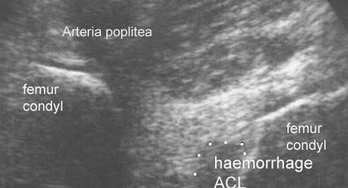
Diagnosis of acute rupture of the anterior cruciate ligament of the knee by sonography.

Skovgaard Larsen LP, Rasmussen OS. Eur J Ultrasound 2000 Dec 1;12(2):163-167.

The accuracy of sonography in the diagnosis of acute rupture of the anterior cruciate ligament (ACL) was tested. Sixty-two patients with a recent traumatic haemarthrosis were examinated. A haematoma at the origin of the ACL in the femoral intercondylar notch was interpreted as evidence of ligament injury. The standard of reference was arthroscopy or clinical follow-up. The sonographic findings were confirmed in 59 of 62 cases. The sensitivity was 88%, the specificity 98%, and the positive and negative predictive values 93 and 96%.

KONDITION

STEP1

GENOPTRÆNING

TRAINING LADDER FOR CHILDREN AND ADOLESCENTS:
FOR RUPTURE OF THE OUTER COLLATERAL LIGAMENT
(RUPTURA COLLATERALE LIGAMENTUM MEDIALE/TIBIALE)

STEP 1

KONDITION
Unlimited: Cycling with raised saddle.

UDSPÆNDING
(5 min)

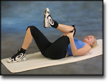
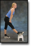
Lie on your back. Draw the injured leg up towards your head so that the muscles in the back of the thigh become increasingly stretched. Perform the exercise with outstretched as well as bent knee. Hold the position for 20 seconds and relax for 20 seconds before repeating. The exercise can also be performed standing with the injured leg outstretched on a chair while the upper body is bent slightly forwards.

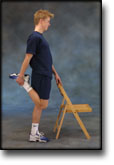
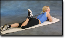
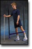
Stand with support from the back of a chair or the wall. Using your hand, bend the knee and draw the foot up and your knee slightly backwards so that the muscles in the front of the thigh become increasingly stretched. Hold the position for 20 seconds and relax for 20 seconds before repeating. The exercise can also be done lying down. If you lie on your stomach you can draw the foot up by using a towel.

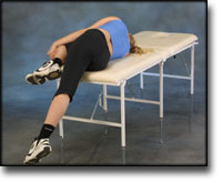
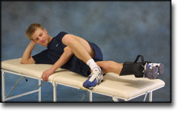
Lie on your side on a table. Bend one leg up under your body and let the other hang over the edge of the table so that the muscles in the outer side of the thigh become increasingly stretched. Hold the position for 20 seconds and relax for 20 seconds before repeating. The exercise can also be done standing by placing the outstretched injured leg behind the good leg at the same time as bending over the injured leg.

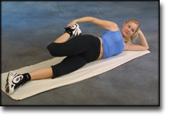
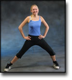
Stand with one leg outstretched and the other slightly bent. Thrust your weight to the side over the bent leg so that the inner side of the opposite thigh becomes increasingly stretched. Hold the position for 20 seconds and relax for 20 seconds before repeating.

KOORDINATION
(5 min)

Seesaw. Balance on two legs, possibly using a hand as support against the wall, balancing subsequently on one leg without support. Look straight ahead and keep knees bent.

STYRKE
(20 min)

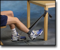
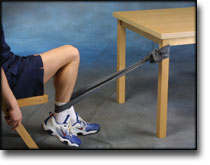
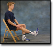
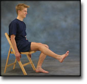
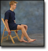
Sit on a chair with elastic around the ankle, facing the elastic. Lift the leg and slowly bend and stretch the knee.

Sit on a chair with elastic attached to the ankle. Raise the leg and slowly stretch and bend the knee.

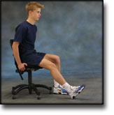
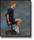
Pull yourself forward on a chair by use of your heel against the floor. Increase the resistance by having someone hold the chair.

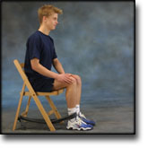
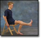
Sit on a chair and lift the knee to a horizontal position. Hold for 1 minute, lower the leg to approx. 45 degrees for 30 seconds. Lower again to the starting position.

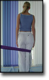
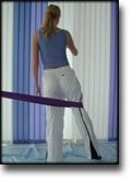
Stand on the healthy leg with the elastic around the inside of the injured leg. Move the injured leg from side to side in a slow smooth movement. Moving the position of the elastic lower down the leg can increase the load.

Stand on the good leg with the elastic around the outer side of the injured leg above the knee. Move the injured leg from side to side in a slow movement.

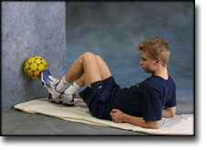
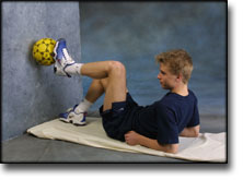
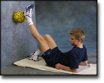
Lie on your back with the healthy leg bent and the injured leg against the wall. Place a ball between the foot and the wall. Move the injured leg up and down the wall while applying a slight pressure on the ball.

Stretching is carried out in the following way: stretch the muscle group for 3-5 seconds. Relax for 3-5 seconds. The muscle group should subsequently be stretched for 20 seconds. The muscle is allowed to be tender, but must not hurt. Relax for 20 seconds, after which the procedure can be repeated.

The time consumed for stretching, coordination and strength training can be altered depending on the training opportunities available and individual requirements.

KONDITION

STEP2

GENOPTRÆNING

TRAINING LADDER FOR CHILDREN AND ADOLESCENTS:
FOR RUPTURE OF THE OUTER COLLATERAL LIGAMENT
(RUPTURA COLLATERALE LIGAMENTUM MEDIALE/TIBIALE)

STEP 2

KONDITION
Unlimited: Cycling. Swimming. Light jogging.

UDSPÆNDING
(5 min)

Lie on your back. Draw the injured leg up towards your head so that the muscles in the back of the thigh become increasingly stretched. Perform the exercise with outstretched as well as bent knee. Hold the position for 20 seconds and relax for 20 seconds before repeating. The exercise can also be performed standing with the injured leg outstretched on a chair while the upper body is bent slightly forwards.

Stand with support from the back of a chair or the wall. Using your hand, bend the knee and draw the foot up and your knee slightly backwards so that the muscles in the front of the thigh become increasingly stretched. Hold the position for 20 seconds and relax for 20 seconds before repeating. The exercise can also be done lying down. If you lie on your stomach you can draw the foot up by using a towel.

Lie on your side on a table. Bend one leg up under your body and let the other hang over the edge of the table so that the muscles in the outer side of the thigh become increasingly stretched. Hold the position for 20 seconds and relax for 20 seconds before repeating. The exercise can also be done standing by placing the outstretched injured leg behind the good leg at the same time as bending over the injured leg.

Stand with one leg outstretched and the other slightly bent. Thrust your weight to the side over the bent leg so that the inner side of the opposite thigh becomes increasingly stretched. Hold the position for 20 seconds and relax for 20 seconds before repeating.

KOORDINATION
(5 min)

Seesaw. Balance on two legs, possibly using a hand as support against the wall, balancing subsequently on one leg without support. Look straight ahead and keep knees bent.

STYRKE
(20 min)

Sit on a chair with elastic around the ankle, facing the elastic. Lift the leg and slowly bend and stretch the knee.

Sit on a chair with elastic attached to the ankle. Raise the leg and slowly stretch and bend the knee.

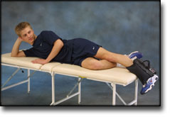
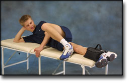
Lie on your side on a mattress or bench. Flex the lower part of the leg and stretch the upper part. It is important to completely stretch the hip. Lift the upper leg upwards with the heel pointing towards the ceiling. Gradually increase the load by attaching a sandbag to the ankle.

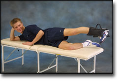
Lie on your side on a mattress or bench. Stretch the lower leg whilst the upper leg is slightly bent. Lift the lower leg stretched upwards. Gradually increase the load by attaching a sandbag to the ankle.

Stand on the healthy leg with the elastic around the inside of the injured leg. Move the injured leg from side to side in a slow smooth movement. Moving the position of the elastic lower down the leg can increase the load.

Stand on the good leg with the elastic around the outer side of the injured leg above the knee. Move the injured leg from side to side in a slow movement.

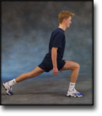
Go forward on the injured leg until the knee is bent to max. 90 degrees. Stand up on the same leg and return to the starting position.

Stand on the injured leg up on a stool with the elastic around your waist. Stand facing the wall. Walk backwards up and down from the stool under resistance from the elastic, alternating between right and left leg first.

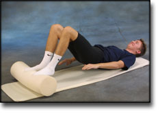
Lie on your back with a ball or firm round cushion under both feet. Roll the ball backwards and forwards in a steady pace while lifting your backside.

Stretching is carried out in the following way: stretch the muscle group for 3-5 seconds. Relax for 3-5 seconds. The muscle group should subsequently be stretched for 20 seconds. The muscle is allowed to be tender, but must not hurt. Relax for 20 seconds, after which the procedure can be repeated.

The time consumed for stretching, coordination and strength training can be altered depending on the training opportunities available and individual requirements.

KONDITION

STEP3

GENOPTRÆNING

TRAINING LADDER FOR CHILDREN AND ADOLESCENTS:
FOR RUPTURE OF THE OUTER COLLATERAL LIGAMENT
(RUPTURA COLLATERALE LIGAMENTUM MEDIALE/TIBIALE)

STEP 3

KONDITION
Unlimited: Cycling. Swimming. Running with increasing speed and cautious directional change.

UDSPÆNDING
(5 min)

Lie on your back. Draw the injured leg up towards your head so that the muscles in the back of the thigh become increasingly stretched. Perform the exercise with outstretched as well as bent knee. Hold the position for 20 seconds and relax for 20 seconds before repeating. The exercise can also be performed standing with the injured leg outstretched on a chair while the upper body is bent slightly forwards.

Stand with support from the back of a chair or the wall. Using your hand, bend the knee and draw the foot up and your knee slightly backwards so that the muscles in the front of the thigh become increasingly stretched. Hold the position for 20 seconds and relax for 20 seconds before repeating. The exercise can also be done lying down. If you lie on your stomach you can draw the foot up by using a towel.

Lie on your side on a table. Bend one leg up under your body and let the other hang over the edge of the table so that the muscles in the outer side of the thigh become increasingly stretched. Hold the position for 20 seconds and relax for 20 seconds before repeating. The exercise can also be done standing by placing the outstretched injured leg behind the good leg at the same time as bending over the injured leg.

Stand with one leg outstretched and the other slightly bent. Thrust your weight to the side over the bent leg so that the inner side of the opposite thigh becomes increasingly stretched. Hold the position for 20 seconds and relax for 20 seconds before repeating.

KOORDINATION
(10 min)

Seesaw. Balance on two legs, possibly using a hand as support against the wall, balancing subsequently on one leg without support. Look straight ahead and keep knees bent.

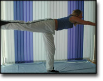
Stand on the injured leg with your upper body bent forwards at 90 degrees. Lift the good leg in a straight line behind you. When you feel comfortable with the exercise, it can be made more difficult by closing your eyes.

STYRKE
(45 min)

Sit on a chair with elastic around the ankle, facing the elastic. Lift the leg and slowly bend and stretch the knee.

Stand on the healthy leg with the elastic around the inside of the injured leg. Move the injured leg from side to side in a slow smooth movement. Moving the position of the elastic lower down the leg can increase the load.

Stand on the healthy leg with the elastic around the outside of the injured leg. Move the injured leg from side to side in a slow smooth movement. Moving the position of the elastic lower down the leg can increase the load.

Lie on your side on a mattress or bench. Flex the lower part of the leg and stretch the upper part. It is important to completely stretch the hip. Lift the upper leg upwards with the heel pointing towards the ceiling. Gradually increase the load by attaching a sandbag to the ankle.

Lie on your side on a mattress or bench. Stretch the lower leg whilst the upper leg is slightly bent. Lift the lower leg stretched upwards. Gradually increase the load by attaching a sandbag to the ankle.

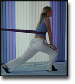
Stand with elastic around the hip. Step forward over one knee and hold the front foot firmly against the floor. Bend the rear leg and go forward onto your toes. Remember to change leg.

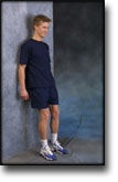
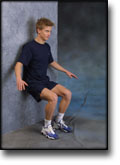
Stand with your back to the wall with your weight on both feet. Slowly go down and bend the knee to 90 degrees, and slowly rise again.

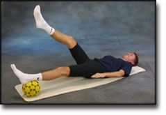
Lie on your back with a ball or firm round cushion under the injured leg. Lift your backside up from the floor and stretch the healthy leg. Hold the position for a few seconds.

Lie on your back with a ball or firm round cushion under both feet. Roll the ball backwards and forwards in a steady pace while lifting your backside.

Stretching is carried out in the following way: stretch the muscle group for 3-5 seconds. Relax for 3-5 seconds. The muscle group should subsequently be stretched for 20 seconds. The muscle is allowed to be tender, but must not hurt. Relax for 20 seconds, after which the procedure can be repeated.

The time consumed for stretching, coordination and strength training can be altered depending on the training opportunities available and individual requirements.