Kategoriarkiv: shinbone

KONDITION

step1

Training ladder for:
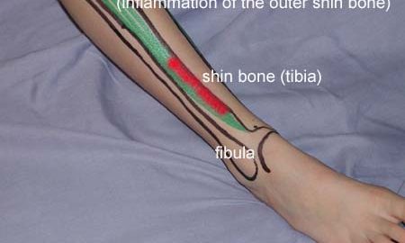
INFLAMMATION OF THE OUTER SHIN BONE
(PERIOSTITIS TIBIALIS LATERALIS)

STEP 1

The indications of time after stretching, coordination training and strength training show the division of time for the respective type of training when training for a period of one hour. The time indications are therefore not a definition of the daily training needs, as the daily training is determined on an individual basis.

KONDITION
Unlimited: Cycling. Swimming. Running in deep water.

UDSPÆNDING
(15 min)

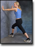
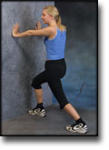
Stand with the injured leg stretched backwards with the toes facing front. Slowly bend the knee so that the calf muscles become increasingly stretched. Hold the position for 20 seconds and relax for 20 seconds before repeating.

Stand with the injured leg outstretched with the ankle joint bent up against the wall. Press your abdomen against the wall so that the calf muscles become increasingly stretched. Hold the position for 20 seconds and relax for 20 seconds before repeating.

Stand on the good leg while gaining support on the wall with the hand on the same side. Bend the knee of the injured leg and draw the heel towards the buttocks. Take hold of the big toe side of the foot and draw slowly upwards so that the ankle joint is stretched to the maximum and drawn slightly outwards so that increased stretching is experienced on the inside of the shin bone. Hold the position for 20 seconds and relax for 20 seconds before repeating.

Stand on the good leg while gaining support from the wall with the opposite hand. Bend the knee of the injured leg and draw the heel towards the buttocks. Take hold of the little toe side of the foot and draw slowly upwards so that the ankle joint is stretched to the maximum and drawn over the good leg so that increased stretching is experienced on the outer side of the shin bone. Hold the position for 20 seconds and relax for 20 seconds before repeating.

KOORDINATION
(5 min)

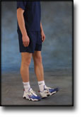
Seesaw. Balance on two legs, possibly using a hand as support against the wall, balancing subsequently on one leg without support. Look straight ahead and keep knees bent.

STYRKE
(40 min)

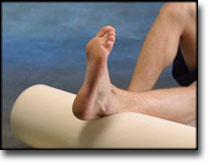
Sit on the floor. Tip the foot up and down with elastic under the forefoot so that the elastic becomes taut.

Stand on both legs. Tip the toes on the leg to be trained upwards and down again, whilst having the heel firmly on the floor during the exercise.

Sit on the floor. Tip the foot from side to side with elastic on the outer side of the foot, without moving the knee.

Sit on the floor. Tip the foot from side to side with elastic on the inner side of the foot, without moving the knee.

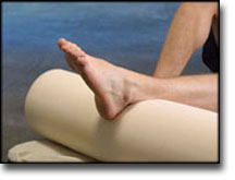
Lay on the floor with a cushion under the calf. Tip the foot up and down without putting any resistance on the foot.

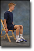
Sit on a chair. Keep the heel firmly on the ground and tip the toes up.

Stretching is carried out in the following way: stretch the muscle group for 3-5 seconds. Relax for 3-5 seconds. The muscle group should subsequently be stretched for 20 seconds. The muscle is allowed to be tender, but must not hurt. Relax for 20 seconds, after which the procedure can be repeated. The time consumed for stretching, coordination and strength training can be altered depending on the training opportunities available and individual requirements.

treatment-article

SportNetDoc

Surgical treatment of medial tibial stress syndrome (shin splint) by fasciotomy of the superficial posterior compartment of the leg.

Holen KJ, Engebretsen L, Grontvedt T, Rossvoll I, Hammer S, Stoltz V. Scand J Med Sci Sports 1995 Feb;5(1):40-3.

From September 1988 to June 1990, 35 athletes were treated for medial tibial stress syndrome (shin splint) by fasciotomy of the superficial posterior compartment of the leg. Thirty-two patients were available for the follow-up, including self-assessment, clinical examination and activity scoring. The mean postoperative observation time was 16 months. Thirteen patients were performing sports at top international or top national level; 19 patients were competing at different lower levels. Twenty-three patients improved, 7 were unchanged and 2 had poor results.

treatment-article

SportNetDoc

Interventions for preventing and treating stress fractures and stress reactions of bone of the lower limbs in young adults.

Gillespie WJ, Grant I. Cochrane Database Syst Rev 2000;(2):CD000450.

BACKGROUND.
Stress reaction in bone, which may proceed to a fracture, is a significant problem in military recruits and in athletes, particularly long distance runners.

OBJECTIVES.
To evaluate the evidence from controlled trials of treatments and programmes for prevention or management of lower limb stress fractures and stress reactions of bone in active young adults.

SEARCH STRATEGY.
We searched the Cochrane Musculoskeletal Injuries Group Trials Register, The Cochrane Library, MEDLINE, EMBASE, Current Contents, Dissertation Abstracts, Index to UK Theses and the bibliographies of identified articles. Date of last search: December 1997.

SELECTION CRITERIA.
Any randomised or quasi-randomised trial evaluating a programme or treatment to prevent or treat lower limb stress reactions of bone or stress fractures in active young adults.

DATA COLLECTION AND ANALYSIS.
Searching, a decision on inclusion or exclusion, methodological assessment, and data extraction were carried out according to a predetermined protocol included in the body of the review. Analysis using Review Manager software allowed pooling of data and calculation of Peto odds ratios and absolute risk reductions, each with 95% confidence intervals.

MAIN RESULTS.
The use of “shock absorbing” insoles, evaluated in four trials, appears to reduce the incidence of stress fractures and stress reactions of bone (Peto odds ratio 0.47, 95% confidence interval 0. 30 to 0.76). Incomplete data from one trial indicated that reduction of running and jumping intensity may also be effective. The use of pneumatic braces in the rehabilitation of tibial stress fractures significantly reduces the time to recommencing training (weighted mean difference -42.6 days, 95% confidence interval -55.8 to -29.4 days).

REVIEWER’S CONCLUSIONS.
The use of shock absorbing insoles in footwear reduces the incidence of stress fractures in athletes and military personnel. Rehabilitation after tibial stress fracture is aided by the use of pneumatic bracing.

examination-article

SportNetDoc

Femoral stress fractures.

Boden BP, Speer KP. Clin Sports Med 1997 Apr;16(2):307-17.

Stress fractures are common overuse injuries attributed to the repetitive trauma associated with vigorous weightbearing activities. A high index of suspicion is necessary to diagnose stress fractures of the femur because the symptoms may be vague. The precipitating factors, whether related to training errors or medical conditions, should be thoroughly evaluated. Early diagnosis of distraction femoral neck stress fractures is critical to avoid serious complications. Femoral shaft stress fractures have excellent healing potential when diagnosed early and treated non-operatively. Stress fractures of the femoral condyles are uncommon, but should be included in the differential of knee pain.

cause-article

SportNetDoc

Risk factors for stress fractures.

Bennell K, Matheson G, Meeuwisse W, Brukner P. Sports Med 1999 Aug;28(2):91-122.

Preventing stress fractures requires knowledge of the risk factors that predispose to this injury. The aetiology of stress fractures is multifactorial, but methodological limitations and expediency often lead to research study designs that evaluate individual risk factors. Intrinsic risk factors include mechanical factors such as bone density, skeletal alignment and body size and composition, physiological factors such as bone turnover rate, flexibility, and muscular strength and endurance, as well as hormonal and nutritional factors. Extrinsic risk factors include mechanical factors such as surface, footwear and external loading as well as physical training parameters. Psychological traits may also play a role in increasing stress fracture risk. Equally important to these types of analyses of individual risk factors is the integration of information to produce a composite picture of risk. The purpose of this paper is to critically appraise the existing literature by evaluating study design and quality, in order to provide a current synopsis of the known scientific information related to stress fracture risk factors. The literature is not fully complete with well conducted studies on this topic, but a great deal of information has accumulated over the past 20 years. Although stress fractures result from repeated loading, the exact contribution of training factors (volume, intensity, surface) has not been clearly established. From what we do know, menstrual disturbances, caloric restriction, lower bone density, muscle weakness and leg length differences are risk factors for stress fracture. Other time-honoured risk factors such as lower extremity alignment have not been shown to be causative even though anecdotal evidence indicates they are likely to play an important role in stress fracture pathogenesis.

treatment-article1

SportNetDoc

Acute exertional compartment syndrome of the medial foot.

Blacklidge DK, Kurek JB, Soto AD, Kissel CG. J Foot Ankle Surg 1996 Jan-Feb;35(1):19-22.

A review of compartment syndrome, both acute and chronic, is presented. The pathophysiology, anatomy, diagnosis, and treatment are presented in relation to a unique case report. The case is one of acute exertional compartment syndrome of the medial foot treated by fasciotomy. This condition is uncommon in both its nature and location.

treatment-article2

SportNetDoc

Acute compartment syndrome due to closed muscle rupture.

Gwynne Jones DP, Theis JC. Aust N Z J Surg 1997 Apr;67(4):227-8.

Acute compartment syndrome has multiple causes: fractures, crush injury, vascular trauma and burns. Exertional compartment syndrome may be acute (progressive) or chronic (usually reversible). The acute form usually occurs after intensive exercise. Closed muscle rupture is an uncommon cause with few reports. We report two cases, in the peroneal compartment of the leg and the flexor compartment of the forearm, to show that a high index of suspicion, allowing prompt diagnosis and fasciotomy, will enable a full recovery without complications.

treatment-article3

SportNetDoc

Acute compartment syndrome.

Engelund D, Kjersgaard AG. Ugeskr Laeger 1991 Apr 15;153(16):1110-3.

The object of this article is to review the current knowledge about the acute compartment syndrome. The syndrome is caused by increased pressure in a muscle compartment and may result from several different conditions: fractures, contusions, haemorrhage, poisoning etc. The pathological physiology is complicated but the main theory is that progressive venous hypertension is involved and that this causes cessation of the microcirculation of the muscle concerned. The clinical diagnosis is described and pressure recording apparatus is reviewed. Treatment of the acute compartment syndrome consists of fasciotomy. Common sites are indicated and operative techniques suggested. Fasciotomy should be performed with compartmental pressures of about 30 mmHg. The untreated compartment syndrome will result in muscular fibrosis and nerve injury and will thus cause incapacitating conditions which may be avoided entirely if fasciotomy is performed in time.