Kategoriarkiv: Thigh, back

KONDITION

step1

Training ladder for:
CHRONIC COMPARTMENT SYNDROME IN THE POSTERIOR THIGH
(KRONISK KOMPARTMENT SYNDROM)

STEP 1

The indications of time after stretching, coordination training and strength training show the division of time for the respective type of training when training for a period of one hour. The time indications are therefore not a definition of the daily training needs, as the daily training is determined on an individual basis.

KONDITION
Unlimited: Cycling. Swimming.

UDSPÆNDING
(20 min)

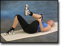
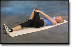
Lie on your back. Draw the injured leg up towards your head so that the muscles in the back of the thigh become increasingly stretched. Perform the exercise with outstretched as well as bent knee. Hold the position for 20 seconds and relax for 20 seconds before repeating. The exercise can also be performed standing with the injured leg outstretched on a chair while the upper body is bent slightly forwards.

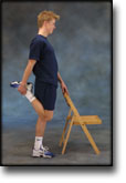
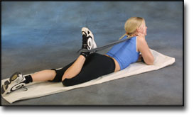
Stand with support from the back of a chair or the wall. Using your hand, bend the knee and draw the foot up and your knee slightly backwards so that the muscles in the front of the thigh become increasingly stretched. Hold the position for 20 seconds and relax for 20 seconds before repeating. The exercise can also be done lying down. If you lie on your stomach you can draw the foot up by using a towel.

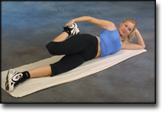
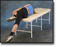
Lie on your side on a table. Bend one leg up under your body and let the other hang over the edge of the table so that the muscles in the outer side of the thigh become increasingly stretched. Hold the position for 20 seconds and relax for 20 seconds before repeating. The exercise can also be done standing by placing the outstretched injured leg behind the good leg at the same time as bending over the injured leg.

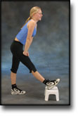
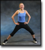
Stand with one leg outstretched and the other slightly bent. Thrust your weight to the side over the bent leg so that the inner side of the opposite thigh becomes increasingly stretched. Hold the position for 20 seconds and relax for 20 seconds before repeating.

Lie on your back with one leg outstretched and the other bent with the foot on the other side of the outstretched leg. Draw the knee up towards the opposite shoulder so that the buttocks become increasingly stretched. Hold the position for 20 seconds and relax for 20 seconds before repeating.

KOORDINATION
(5 min)

Seesaw. Balance on two legs, possibly using a hand as support against the wall, balancing subsequently on one leg without support. Look straight ahead and keep knees bent.

STYRKE
(40 min)

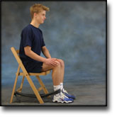
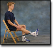
Sit on a chair with elastic attached to the ankle. Raise the leg and slowly stretch and bend the knee.

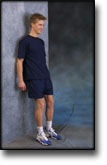
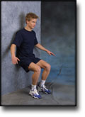
Stand with your back to the wall with your weight on both feet. Slowly go down and bend the knee to 90 degrees, and slowly rise again.

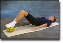
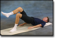
Lie on your back with a ball or firm round cushion under both feet. Raise your backside up from the floor and hold your feet on the ball. Hold the position for a few seconds.

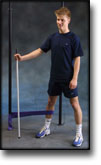
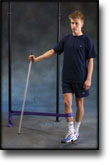
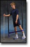
Stand on the healthy leg with the elastic around the inside of the injured leg. Move the injured leg from side to side in a slow smooth movement. Moving the position of the elastic lower down the leg can increase the load.

Stand on the healthy leg with the elastic around the outside of the injured leg. Move the injured leg from side to side in a slow smooth movement. Moving the position of the elastic lower down the leg can increase the load.

Lie on your back and bend the injured leg. Lift the healthy leg, and then raise your backside with the weight on the injured leg to be trained. Hold the position a few seconds and lower your backside again. Moving the injured leg further away from the body will increase the load.

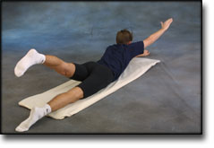
Lie on your stomach on the floor with your arms above your head and with outstretched legs. Lift right arm and left leg together, changing to lift left arm and right leg together.

Stretching is carried out in the following way: stretch the muscle group for 3-5 seconds. Relax for 3-5 seconds. The muscle group should subsequently be stretched for 20 seconds. The muscle is allowed to be tender, but must not hurt. Relax for 20 seconds, after which the procedure can be repeated. The time consumed for stretching, coordination and strength training can be altered depending on the training opportunities available and individual requirements.

treatment-article3

SportNetDoc

Good results of fasciotomy in chronic compartment syndrome of the lower leg.

Verleisdonk EJ, van den Helder CJ, Hoogendoorn HA, van der Werken C. Ned Tijdschr Geneeskd 1996 Dec 14;140(50):2513-7.

OBJECTIVE.
To assess the results of fasciotomy in patients with a chronic compartmental syndrome. DESIGN: Retrospective study.

SETTING.
Department of Surgery, Central Military Hospital, Utrecht, the Netherlands. METHOD: Closed fasciotomy was performed in 81 patients (151 compartments) after standardized measurement of the pressure of the symptomatic compartment during exercise. The anterior compartment was affected 149 times and the lateral compartment twice. The pressure reading was repeated at least 3 months after the operation. All operated patients 6 months postoperatively were sent a written questionnaire inquiring about the results of the operation.

RESULTS.
Postoperative complications included a neurinoma (3 times) and a seroma (once). The mean postoperative intramuscular pressures were lower than the preoperative ones: the pressure at rest fell from 22.1 to 14.0 mm Hg (p < 0.05), the exercise pressure from 57.5 to 25.4 mm Hg (p < 0.01) and the relaxation pressure from 34.4 to 25.2 mm Hg (p < 0.05). Tn patients had an unchanged increased pressure after the operation, for which a second fasciotomy was performed 4 times. Attenuation of symptoms was reported by 59 patients (76%). Nine patients with poor results had already had a combination with some other hyperpressure injury before the operation.

CONCLUSION.
Closed fasciotomy in a demonstrated chronic compartmental syndrome in most cases gave good results, viz. attenuation of symptoms and a decrease of the intramuscular pressure, especially after exercise.

treatment-article2

SportNetDoc

Chronic compartment syndrome of the quadriceps femoris muscle in athletes. Diagnosis, imaging and treatment with fasciotomy.

Orava S, Laakko E, Mattila K, Makinen L, Rantanen J, Kujala UM. Ann Chir Gynaecol 1998;87(1):53-8.

BACKGROUND AND AIMS.
Chronic quadriceps femoris muscle compartment syndrome is described.

MATERIAL AND METHODS.
Over a 13 year period nine patients were diagnosed, treated surgically by fasciotomy and followed up at a referral center specialized in sports traumatology. There were four power lifters, three body builders, one endurance walker and one cyclist in the series. The use of anabolic steroids was admitted by four of the nine patients. The patients complained of gradually worsening pain on the anterolateral side of the thigh during training. Initially the pain appeared only at the end of the training session. As the syndrome became more severe, the pain began earlier during exercise and gradually became worse preventing maximal training. Skeletal radiographs and ultrasound examinations were normal, except in two patients, who had a positive echography finding with local atrophy. MRI examination confirmed the US diagnosis.

RESULTS AND CONCLUSIONS.
As conservative treatment did not provide relief of symptoms, a fasciotomy anterior to the iliotibial tract was performed bilaterally to seven patients and unilaterally to two patients. Biopsies from the atrophied sites showed muscle cell necrosis. All the patients recovered well after the surgery and were able return to their original level of sport. The only complication of surgery was a postoperative hematoma in one patient, which delayed the beginning of the training, but did not result in any persistent complaints.

treatment-article1

SportNetDoc

Treatment of chronic exertional anterior compartment syndrome with massage: a pilot study.

Blackman PG, Simmons LR, Crossley KM. Clin J Sport Med 1998 Jan;8(1):14-7.

OBJECTIVE.
To determine the effect of massage on anterior chronic exertional compartment syndrome (CECS) with respect to symptoms, intracompartmental pressures, and work output of the anterior compartment in dorsiflexion.

DESIGN.
One group-repeated measures design.

SETTING.
A private sports medicine clinic in Melbourne, Australia.

PARTICIPANTS.
Seven athletes (six men and one woman), aged between 21 and 29 years, were selected on the basis of clinical suspicion for anterior CECS. Historical questionnaire and examination were followed by intracompartmental pressure testing of the anterior compartment. Study exclusion criteria were history of a bleeding diathesis and previous treatment consisting of compartment fasciotomy or massage. All athletes completed the study.

INTERVENTIONS.
A 5-week course of massage consisting of two sessions in the first week and one session per week thereafter, for a total of six treatments. Between each session, a twice-daily standard stretching program involving both anterior and posterior compartments was performed.

MAIN OUTCOME MEASURES.
Postexercise anterior compartment pressures (mm Hg) before and after treatment, work output (J) in dorsiflexion to pain onset before and after treatment, self-reported symptoms before and after treatment.

RESULTS.
There was no significant difference in the 3-minute postexercise compartment pressures after the treatment. There was a significant (p = 0.016) increase, however, in work performed in dorsiflexion to pain onset following the massage course.

CONCLUSIONS.
Intermittent massage combined with specific stretching should be considered in the treatment of anterior CECS.

examination-article

SportNetDoc

Intracompartmental pressure before and after fasciotomy in runners with chronic deep posterior compartment syndrome.

Biedert RM, Marti B. Int J Sports Med 1997 Jul;18(5):381-6.

Exercise induced pain in the posterior part of the leg is common among runners; the underlying reason for these complaints may be very different. The purpose of the present, controlled study was therefore 1. to confirm a clinically diagnosed deep posterior compartment syndrome by using intramuscular pressure measurements and 2. to evaluate the effect of a surgical release on clinical signs and intracompartment pressure values. Fifteen symptomatic runners with the clinical suspicion of a chronic deep posterior compartment syndrome and nine healthy recreational runners as controls were investigated. Intramuscular pressure was measured both at rest and up to two minutes post-exercise, using a pressure-monitor with a transducer. In symptomatic runners, the average pressure was preoperatively 5.6 mmHg (95%-confidence-interval [CI]: 3.4-7.6) at rest, rising to 18.5 mmHg (CI: 15.4-21.8) post-exercise. Corresponding values in healthy control runners were 5.1 mmHg (CI: 1.9-8.3) at rest, with a decrease induced by exercise to 2.8 mmHg (CI: -0.5-6.1). After fasciotomy of the deep posterior compartment in all fifteen symptomatic runners, average pressure values fell to 2.2 mmHg (CI: 1.0-3.4) at rest, and were further reduced after (now pain-free) exercise to 1.6 mmHg (CI: 0.6-2.6). The decrease between pre-operative and post-operative values was statistically highly significant (p < 0.0001 for values after running, p < 0.005 for values at rest). In conclusion, intracompartment pressure measurement is a useful technique to confirm the clinical diagnosis of deep posterior compartment syndrome prior to recommending surgery. Hereby, an exercise-induced rise in pressure of at least 10 mmHg, corresponding to a two- to threefold increase of values measured at rest, may be a more important diagnostic criterion than absolute levels of pressure measured before or after running.

Rehabilitation-article

SportNetDoc

Femoral shaft stress fractures in athletes.

Hershman EB, Lombardo J, Bergfeld JA. Clin Sports Med 1990 Jan;9(1):111-9.

Stress fractures of the femoral shaft in athletes occur most commonly in the proximal third of the femur. They can, however, also be found in the mid- or distal third. Conservative treatment is highly successful in healing these fractures without complications. Athletes can usually return to activity in 8 to 14 weeks. Recognition of the symptoms characteristic of these fractures (vague thigh pain, diffuse tenderness, no trauma) will assist early diagnosis. Early definitive diagnosis can be made by radionuclide scanning or later, by plain radiography, if symptoms have been present for a sufficient period. Diagnosis is not limited to novice runners since runners with significant mileage, or baseball or basketball players, can develop femoral shaft stress fractures.

treatment-article

SportNetDoc

Interventions for preventing and treating stress fractures and stress reactions of bone of the lower limbs in young adults.

Gillespie WJ, Grant I. Cochrane Database Syst Rev 2000;(2):CD000450.

BACKGROUND.
Stress reaction in bone, which may proceed to a fracture, is a significant problem in military recruits and in athletes, particularly long distance runners.

OBJECTIVES.
To evaluate the evidence from controlled trials of treatments and programmes for prevention or management of lower limb stress fractures and stress reactions of bone in active young adults.

SEARCH STRATEGY.
We searched the Cochrane Musculoskeletal Injuries Group Trials Register, The Cochrane Library, MEDLINE, EMBASE, Current Contents, Dissertation Abstracts, Index to UK Theses and the bibliographies of identified articles. Date of last search: December 1997.

SELECTION CRITERIA.
Any randomised or quasi-randomised trial evaluating a programme or treatment to prevent or treat lower limb stress reactions of bone or stress fractures in active young adults.

DATA COLLECTION AND ANALYSIS.
Searching, a decision on inclusion or exclusion, methodological assessment, and data extraction were carried out according to a predetermined protocol included in the body of the review. Analysis using Review Manager software allowed pooling of data and calculation of Peto odds ratios and absolute risk reductions, each with 95% confidence intervals.

MAIN RESULTS.
The use of “shock absorbing” insoles, evaluated in four trials, appears to reduce the incidence of stress fractures and stress reactions of bone (Peto odds ratio 0.47, 95% confidence interval 0. 30 to 0.76). Incomplete data from one trial indicated that reduction of running and jumping intensity may also be effective. The use of pneumatic braces in the rehabilitation of tibial stress fractures significantly reduces the time to recommencing training (weighted mean difference -42.6 days, 95% confidence interval -55.8 to -29.4 days).

REVIEWER’S CONCLUSIONS.
The use of shock absorbing insoles in footwear reduces the incidence of stress fractures in athletes and military personnel. Rehabilitation after tibial stress fracture is aided by the use of pneumatic bracing.

examination-article2

SportNetDoc

Femoral stress fractures.

Boden BP, Speer KP. Clin Sports Med 1997 Apr;16(2):307-17.

Stress fractures are common overuse injuries attributed to the repetitive trauma associated with vigorous weightbearing activities. A high index of suspicion is necessary to diagnose stress fractures of the femur because the symptoms may be vague. The precipitating factors, whether related to training errors or medical conditions, should be thoroughly evaluated. Early diagnosis of distraction femoral neck stress fractures is critical to avoid serious complications. Femoral shaft stress fractures have excellent healing potential when diagnosed early and treated non-operatively. Stress fractures of the femoral condyles are uncommon, but should be included in the differential of knee pain.

examination-article1

SportNetDoc

Stress fractures of the femoral shaft in athletes–more common than expected. A new clinical test.

Johnson AW, Weiss CB Jr, Wheeler DL. Am J Sports Med 1994 Mar-Apr;22(2):248-56.

Athletes from 20 Division I AA collegiate varsity sports and 1 club sport were followed carefully for the development of stress fractures during the 1990 to 1991 and the 1991 to 1992 academic years. During this period, among 914 athletes, 34 stress fractures were sustained. Seven of these, or 20.6%, were of the femoral shaft. This represents a much higher incidence than previously observed in athletes. A new clinical test is described that significantly aids in the early diagnosis and follow-up treatment of femoral shaft stress fractures.