Kategoriarkiv: Thigh

article1.2

SportNetDoc

Myositis ossificans of the upper extremity: a long-term follow-up.

Carlson WO, Klassen RA. J Pediatr Orthop 1984 Nov;4(6):693-6.

The long-term results of nonoperative treatment of myositis ossificans traumatica have been infrequently reported. We reviewed 83 cases of myositis ossificans seen at the Mayo Clinic from 1950 to 1979 in patients up to 21 years of age. The upper extremity was involved in 31 patients. Follow-up averaged 13 years for 23 of the (74%) patients studied. Football injuries had occurred in 20 of these patients, and the diagnosis was made an average of 3.3 weeks from the time of the initial injury. Observation was the only treatment in 18 of the 31 cases. At follow-up two-thirds of the patients with upper extremity involvement had no problems associated with the disorder, and one-third described some difficulties. Nonoperative treatment remains an accepted management approach for this problem.

article1.1

SportNetDoc

Treatment of traumatic myositis ossificans circumscripta; use of aspiration and steroids.

Molloy JC, McGuirk RA. J Trauma 1976 Nov;16(11):851-7.

The classification, clinical presentation, forms of treatment, and theories of pathogenesis of traumatic myositis ossificans circumscripta are reviewed. A group of 7 patients with this lesion was treated with hematoma aspiration and injection of steroids, lysosomal enzymes, and xylocaine, resulting in significant limitation of disability. The determining factor in the development of this disorder seems to be the response of local soft tissues to trauma rather than the magnitude of the trauma itself which we feel accounts for the success of the form of treatment described.

complications for muscle ruptures


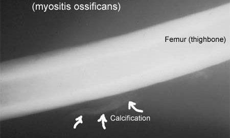
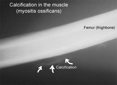
  1. Calcification in the muscle (Myositis ossificans):
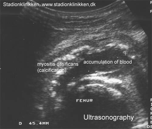
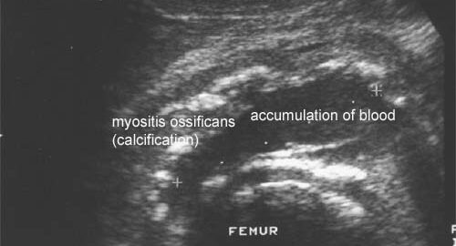
    Muscles which have been subjected to a rupture can from time to time be found to have some calcification, although the reasons for this are unknown. The diagnosis is often made due to the rehabilitation not progressing as expected in relation to the primary evaluation of the extent of the injury. The calcification can be seen under ultrasound scanning after a few days, and subsequently on an x-ray after a few weeks (X-ray picture), (Ultrasonic image) . A considerably longer rehabilitation period must be expected if the muscular bleeding is complicated by calcification in the muscle. Surgery is almost never indicated, as an operation can involve further calcification. Vigorous massage on the blood accumulation should be avoided on the same grounds (article-1), (article-2).
  2. Formation of scar tissue in the muscles (cicatricial tissue).
    Formation of scar tissue is often seen after a muscle rupture, and can in some cases cause permanent discomfort. The formation of scar tissue is often the cause of the rehabilitation progressing slower than anticipated, as well as relapses a long time after the injury initially occurring. A local tenderness in the muscle can usually be experienced, aggravated when activating and stretching the muscle. It is often possible to detect scar tissue under ultrasound scanning, however, this can be quite difficult and requires a doctor well experienced in performing ultrasound scans. An area of inflammation will often be seen surrounding the scar tissue formation.
  3. Blood accumulation in the muscle (haematoma musculi).
    Injuries to muscles always involve muscular bleeding to a greater or lesser extent. In some cases the bleeding is diffuse between the muscle cells, whereas other cases can involve the bleeding being a larger, well-defined accumulation in the muscle. The larger the accumulation, the longer period is required for the accumulation to disappear and allow the muscle to heal. Accumulations of blood in the muscles often entail more pronounced pain than anticipated in relation to the primary evaluation of the extent of the injury. The diagnosis is best made via use of ultrasound scanning. If the accumulation is large it can be drained, which can advantageously be performed under guidance of ultrasound.
    Some recommend advise caution regarding massage to reduce the risk of myositis ossificans.
  4. Formation of fluid in the muscle (Hygrom).
    A formation of fluid will sometimes appear after an injury to a muscle, and can be of a considerable size. If the fluid does not diminish following relief, the accumulation can be drained by use of ultrasound scanning. Fluid accumulations can be the cause of the rehabilitation not progressing smoothly.
  5. Acute compartment syndrome:
    The groups of muscles on the arms and legs are surrounded by taught muscle membranes (fascias), which are partially unremitting. Damage to blood vessels and nerves can be caused if the bleeding and fluid accumulation in the muscles reaches such a size that the pressure in the muscle group increases. Serious damage to muscles can be caused in acute cases which result in the blood vessels closing. The symptoms comprise increasing pain in the damaged arm or leg muscle, which is often more powerful than expected from the primary evaluation of the extent of the injury. At the same time sensory disturbances can occur. The diagnosis is made by performing a pressure measurement in the muscle compartment, (article). The treatment comprises acute severing of the muscle membrane. It is imperative for the continuing function of the muscle that this operation will be acute, which is, of course, only possible if the athlete seeks acute medical attention, (article-1), (article-2), (article-3).
  6. Chronic compartment syndrome:
    The groups of muscles on the arms and legs are surrounded by taught muscle membranes (fascias), which are partially unremitting. Pain can be experienced in a muscle group in the leg after only a few minutes activity following an earlier muscle injury, or following a very rapid increase in training of individual muscle groups. There is a sensation that the muscle is “tightened” and becomes hard, which is accompanied by discomfort. If the activity is stopped the discomfort diminishes, but returns a short period after the resumption of sports activity. This can be due to the muscle swelling up (after injury), or growing quicker than the muscle membrane can manage to keep up to (increase in training intensity too fast), whereby the pressure on the blood vessels and nerves in the muscle group increases. The diagnosis can be made with a pressure measurement in the muscle compartment, (article). Treatment comprises relief with slowly increasing training intensity after loss of symptoms. In cases where there is a lack of progress a surgical severing of the muscle membranes can be performed, which is usually a minor procedure with good results. (article-1), (article-2).

KONDITION

STEP4

GENOPTRÆNING

TRAINING LADDER FOR CHILDREN AND ADOLESCENTS:
FOR MUSCLE RUPTURE IN THE ANTERIOR THIGH
(RUPTURA MUSCULI)

STEP 4

The following rehabilitation program will cover the needs for the vast majority of children with muscle rupture in the anterior thigh. Older teenagers involved in sports at a high level can advantageously use the rehabilitation program for adults.
KONDITION
Unlimited: Cycling. Swimming. Running with jumping.

UDSPÆNDING
(5 min)

Stand with support from the back of a chair or the wall. Using your hand, bend the knee and draw the foot up and your knee slightly backwards so that the muscles in the front of the thigh become increasingly stretched. Hold the position for 20 seconds and relax for 20 seconds before repeating. The exercise can also be done lying down. If you lie on your stomach you can draw the foot up by using a towel.

STYRKE
(15 min)

Sit on a chair with elastic attached to the ankle. Raise the leg and slowly stretch and bend the knee.

Lie on the floor with slightly bent knees. Put the elastic under the foot and hold firmly with your hands. Stretch the knee so that the elastic is drawn tight and slowly bend the knee again.

Stand with your back against a wall with a ball or firm round cushion between the wall and your back. Slowly go down to bend your knee 90 degrees before slowly rising up again.

Stand with elastic around the hip. Step forward over one knee and hold the front foot firmly against the floor. Bend the rear leg and go forward onto your toes. Remember to change leg.

Stand with both legs on the stool with elastic around the hip. Take-off and land with feet together.

Stretching is carried out in the following way: stretch the muscle group for 3-5 seconds. Relax for 3-5 seconds. The muscle group should subsequently be stretched for 20 seconds. The muscle is allowed to be tender, but must not hurt. Relax for 20 seconds, after which the procedure can be repeated.

The time consumed for stretching, coordination and strength training can be altered depending on the training opportunities available and individual requirements.

KONDITION

STEP3

GENOPTRÆNING

TRAINING LADDER FOR CHILDREN AND ADOLESCENTS:
FOR MUSCLE RUPTURE IN THE ANTERIOR THIGH
(RUPTURA MUSCULI)

STEP 3

The following rehabilitation program will cover the needs for the vast majority of children with muscle rupture in the anterior thigh. Older teenagers involved in sports at a high level can advantageously use the rehabilitation program for adults.
KONDITION
Unlimited: Cycling. Swimming. Running with increasing distance.

UDSPÆNDING
(5 min)

Stand with support from the back of a chair or the wall. Using your hand, bend the knee and draw the foot up and your knee slightly backwards so that the muscles in the front of the thigh become increasingly stretched. Hold the position for 20 seconds and relax for 20 seconds before repeating. The exercise can also be done lying down. If you lie on your stomach you can draw the foot up by using a towel.

STYRKE
(15 min)

Sit on a chair with elastic attached to the ankle. Raise the leg and slowly stretch and bend the knee.

Lie on the floor with slightly bent knees. Put the elastic under the foot and hold firmly with your hands. Stretch the knee so that the elastic is drawn tight and slowly bend the knee again.

Stand with your back to the wall with your weight on both feet. Slowly go down and bend the knee to 90 degrees, and slowly rise again.

Go forward on the injured leg until the knee is bent to max. 90 degrees. Stand up on the same leg and return to the starting position.

Stretching is carried out in the following way: stretch the muscle group for 3-5 seconds. Relax for 3-5 seconds. The muscle group should subsequently be stretched for 20 seconds. The muscle is allowed to be tender, but must not hurt. Relax for 20 seconds, after which the procedure can be repeated.

The time consumed for stretching, coordination and strength training can be altered depending on the training opportunities available and individual requirements.

KONDITION

STEP2

GENOPTRÆNING

TRAINING LADDER FOR CHILDREN AND ADOLESCENTS:
FOR MUSCLE RUPTURE IN THE ANTERIOR THIGH
(RUPTURA MUSCULI)

STEP 2

The following rehabilitation program will cover the needs for the vast majority of children with muscle rupture in the anterior thigh. Older teenagers involved in sports at a high level can advantageously use the rehabilitation program for adults.
KONDITION
Unlimited: Cycling with a weak load. Swimming. Jogging.

UDSPÆNDING
(5 min)

Stand with support from the back of a chair or the wall. Using your hand, bend the knee and draw the foot up and your knee slightly backwards so that the muscles in the front of the thigh become increasingly stretched. Hold the position for 20 seconds and relax for 20 seconds before repeating. The exercise can also be done lying down. If you lie on your stomach you can draw the foot up by using a towel.

STYRKE
(10 min)

Sit on a chair with elastic attached to the ankle. Raise the leg and slowly stretch and bend the knee.

Go up and down from the stool. Go up with alternating right and left legs.

Lie on the floor with slightly bent knees. Put the elastic under the foot and hold firmly with your hands. Stretch the knee so that the elastic is drawn tight and slowly bend the knee again.

Stretching is carried out in the following way: stretch the muscle group for 3-5 seconds. Relax for 3-5 seconds. The muscle group should subsequently be stretched for 20 seconds. The muscle is allowed to be tender, but must not hurt. Relax for 20 seconds, after which the procedure can be repeated.

The time consumed for stretching, coordination and strength training can be altered depending on the training opportunities available and individual requirements.

STEP1

Diagnosis: MUSCLE RUPTURE IN THE ANTERIOR THIGH
(RUPTURA MUSCULI)


Anatomy:
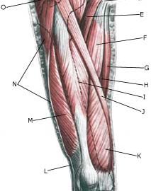
A rupture can in principle occur to all muscles in the thigh, however, ruptures most often happen in the anterior muscle (M quadriceps femoris) which has the function of stretching the knee and flexing the hip. The anterior thigh muscle consists of four muscles (M vastus lateralis, M vastus medialis, M rectus femoris and the deep lying M vastus intermedius).

  1. Spina iliaca anterior superior
  2. M. iliopsoas
  3. Lig. inguinale
  4. Tuberculum pubicum
  5. M. pectineus
  6. M. adductor longus
  7. M. gracilis
  8. M. adductor magnus
  9. M. rectus femoris
  10. M. sartorius
  11. M. vastus medialis
  12. Tractus iliotibialis
  13. M. vastus lateralis
  14. M. tensor fasciae latae et tractus iliotibialis
  15. M. gluteus medius

THIGH FROM THE FRONT

Cause: When a muscle is subjected to a load beyond the strength of the muscle (jump, kick), a rupture occurs. The vast majority of ruptures are partial muscle ruptures. The weakest point is often at the junction between the muscle tendon and the muscle belly. Muscle ruptures in children and adolescents are relatively rare compared with adults.

Symptoms: In slight cases a local tenderness is felt after being subjected to load (“sprained muscle”, “imminent pulled muscle”). In severe cases sudden shooting pains are felt in the muscle (“partial muscle rupture”, “pulled muscle”) and in the worst case a sudden snap is felt rendering the muscle unusable (“total muscle rupture”). The following three symptoms are characteristic in connection with muscle injuries: pain upon applying pressure, stretching and activation of the muscle (stretching knee) against resistance. With total ruptures a defect can often be seen and felt in the muscle, and above and below the rupture a swelling can be felt (the contracted muscle belly and bleeding). The most frequent place for partial ruptures on the anterior thigh is approximately 10 cm below the upper front iliac crest projection (spina iliaca anterior superior) in the rectus femoris muscle.

Acute treatment: Click here.

Examination: In very slight cases (light muscle sprains) with only minimal tenderness and no discomfort when walking normally, medical examination is not necessarily required. The severity of the tenderness is however, not always a measure of the extent of the injury. In cases of more pronounced tenderness or pain, medical examination is required to ensure the diagnosis and treatment. The diagnosis is usually made following normal medical examination, however, if there is any doubt concerning the diagnosis, ultrasound scanning can be performed, as it is the most suitable examination to ensure the diagnosis (Ultrasonic image). The larger the bleeding as assessed by ultrasound scanning, the longer the period needed to heal the injury (article).

Treatment: The treatment of the vast majority of muscle injuries today involves relief and rehabilitation. It is only in very rare cases that surgery is indicated (e.g. total rupture in the anterior muscle tendon close to the attachment on the upper knee cap where surgery is recommended very quickly (article). Even large ruptures in the thigh muscles will usually be able to be healed and rehabilitated without giving functional disorder (but often cosmetic disfigurement with an irregular thigh muscle).

Rehabilitation of children and adolescents: INSTRUCTION

Complications: If steady progress is not experienced, you should be medically (re)examined to ensure that the diagnosis is correct or whether complications for muscle ruptures have arisen.

treatment-article1

SportNetDoc

Complete quadriceps tendon ruptures.

Rougraff BT, Reeck CC, Essenmacher J. Orthopedics 1996 Jun;19(6):509-14.

Forty-four patients with 53 quadriceps tendon ruptures were studied retrospectively with an average follow up of 67.2 months (range: 24 to 155). The type of repair was not associated with differences in functional outcome, patient satisfaction, range of motion, or isokinetic testing. Patients with a delay in surgical treatment in comparison to those immediately repaired had significantly worse functional results (P < .05), lower satisfaction scores (P < .05), and lower isokinetic data for both the injured and uninjured extremities, however, their range of motion and comparative extensor power results were nearly identical. On reviewing all patients, the ultimate range of motion was within an average of 2 degrees of the uninjured side in nearly all patients. Based on the results of this study, all surgical methods can be expected to give comparable results as long as treatment is begun within 1 week of the injury. Regaining range of motion comparable to the uninjured side was not a problem in this series despite a delay in treatment.