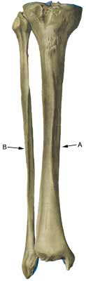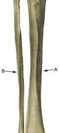
STRESS FRACTURE
|
||
|
||
| Cause: Repeated uniform loads, particularly when walking or running, can cause such great stress that cracks (fractures) appear in the bone (article).
Symptoms: Pain upon applying pressure (direct and indirect tenderness) and applying load (walking, running). Examination: X-ray. Since many stress fractures are not visible early in the course, x-ray examination can be repeated after a few weeks, if stress fractures are still suspected. Scintigraphy, MRI and ultrasound scans can often diagnose stress fractures far earlier than x-rays (Ultrasonic image) (Photo). It is crucial for the result of the treatment that the diagnosis is made as early as possible (article). Treatment: The treatment primarily comprises relief and possibly bandaging. Only in special cases is surgery necessary. It is imperative that there are good shock-absorbing soles in the shoes (article). Rehabilitation: The rehabilitation is completely dependant on the type of fracture and the treatment (relief or surgical). Complications: If progress is not smooth, you should be medically re-evaluated to ensure that the fracture is healing according to plan. In some cases a false joint is formed (pseudoarthrosis), which requires surgical treatment. Special: Shock absorbing shoes or inlays will reduce the load. |