|
||
|
||
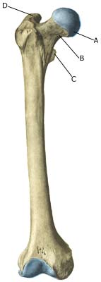
| Cause: Fracture of the thigh bone occurs most commonly following a heavy blow or twist. By far the majority of fractures occur in the middle section of the thigh bone. A fracture of the femoral neck (article) and stress fractures are very seldom seen in children (article).
Symptoms: Pain and swelling. It will most often be impossible for the patient to support himself on the leg due to pain. Treatment: The treatment primarily comprises relief (article). Only in special cases is surgery necessary (article). Rehabilitation of children and adolescents: The rehabilitation is completely dependant on the severity of the fracture and the treatment. All rehabilitation should therefore be performed in close cooperation with the doctor controlling the treatment. A period of at least two months is usually recommended before full participation in sport can be permitted. Complications: The great majority of cases heal without complication or after-effects following non-operative treatment. Complications are more frequently seen following surgical treatment of the fracture (article). A shortening of the leg can be seen following a thigh bone fracture (article) and problems in the healing process where in some cases a false joint is formed (pseudoarthrosis), which requires surgical treatment. |


