Alle indlæg af urtehave_com

KONDITION

step1

Training ladder for:
MUSCLE INFILTRATIONS IN THE NECK/SHOULDER
(MYOSER)

STEP 1

The indications of time after stretching, coordination training and strength training show the division of time for the respective type of training when training for a period of one hour. The time indications are therefore not a definition of the daily training needs, as the daily training is determined on an individual basis.

KONDITION
Unlimited: Cycling. Swimming. Running.

UDSPÆNDING
(20 min)

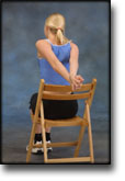
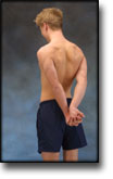
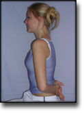
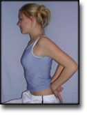
Sit on a chair with your arms outstretched behind your back with hands together. Lift your arms up and backwards so that the front of the shoulders becomes increasingly stretched. Hold the position for 20 seconds and relax for 20 seconds before repeating.

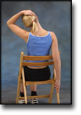
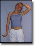
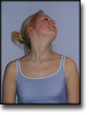
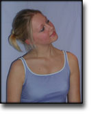
Sit on a chair with one hand on your head and the other holding the chair seat. Slowly draw your head to the side while resisting with the opposite arm so that the muscles on the side of the neck become increasingly stretched. Draw your head in different directions so that all the muscles around the neck are stretched. Hold the position for 20 seconds and relax for 20 seconds before repeating.

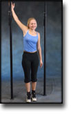
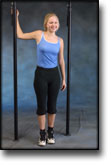
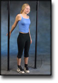
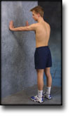
Stand in a doorframe. Press your arms against the frame so that the front of your shoulders become increasingly stretched. Move your arms up and down the doorframe so that different parts of your muscles are stretched. Hold the position for 20 seconds and relax for 20 seconds before repeating.

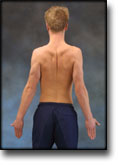
Stand with your hands together behind your back. Draw your shoulder blades together (imagine trying to hold a pencil between your shoulder blades). Hold the position for 10 seconds and rest for 10 seconds before repeating.

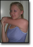
Stand with the injured arm in front of your body. With the opposite hand, press the elbow of the injured arm towards the opposite shoulder, so that the upper part of the arm and the outer shoulder experiences increased stretching. Hold the position for 20 seconds and relax for 20 seconds before repeating.

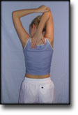
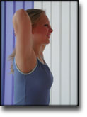
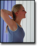
Bend your injured arm behind your head and using the opposite hand, pull the elbow of the injured arm towards the opposite shoulder so that you feel increased stretching. Hold the position for 20 seconds and relax for 20 seconds before repeating.

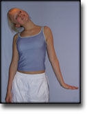
Stand with right arm outstretched down by your side. Turn your hand inwards so that the thumb faces backwards, and move your arm back and down. Pull your chin in and bend your head the opposite way. Using your left hand, gently pull on the back of your head so that the neck and shoulder muscles on the right side become increasingly stretched. Hold the position for 20 seconds and relax for 20 seconds. Repeat the exercise for the opposite side.

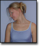
Stretch your left arm down and outwards 30 degrees while bending the wrist upwards. Bend your head the opposite way and turn your chin upwards so that the neck and shoulder muscles on the left side become increasingly stretched. Hold the position for 20 seconds and relax for 20 seconds. Repeat the exercise for the opposite side.

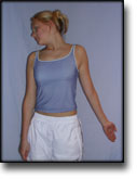
Stretch your left arm down, back and outwards 30 degrees. Bend your head the opposite way and look down your shoulder so that the neck and shoulder muscles on the left side become increasingly stretched. Hold the position for 20 seconds and relax for 20 seconds. Repeat the exercise for the opposite side.

Sit on a chair holding the back of your hands under the lower part of the back. Press your elbows forwards and inwards so that the muscles on the back of the shoulder joint become increasingly stretched. Hold the position for 20 seconds and follow by pressing the elbows backwards so that increased stretching occurs on the front of the shoulder joint. Hold the position for 20 seconds.

STYRKE
(40 min)

Sit on a chair with your hands behind your head. Keeping your chin level, press your head into your hands. Hold the position for 5 seconds. Turn your head upwards and backwards towards the left. Hold the position for 5 seconds. Repeat the exercise for the opposite side.

Turn your head down and to the right. Hold the pressure for 5 seconds before turning your head up, backwards and to the left. Hold the pressure for 5 seconds. Repeat the exercise for the opposite side.

Bend your head so that your ear is close to your shoulder and draw your chin in. Hold the pressure for 5 seconds. Turn your head the other way and hold the pressure for 5 seconds.

Support with both hands against a wall. Put your weight on your hands and draw your shoulder blades backwards and forwards.

Stand with your arms outstretched at your sides with the palms of your hands facing forwards. Draw your shoulder blades together (imagine trying to hold a pencil between your shoulder blades). Hold the position for 10 seconds and rest for 10 seconds before repeating.

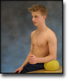
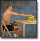
Sit at a table with the injured arm’s elbow on a ball. Press against the ball with a slow movement for 5 seconds. Rest for 5 seconds before repeating.

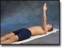
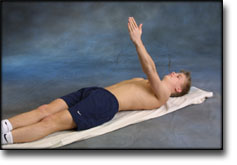
Lie on your back with the injured arm pointing upwards. Stretch the arm further up so that the shoulder blade lifts from the floor, and go down again. The arm must be outstretched the whole time.

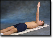
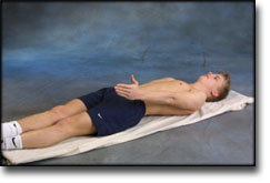
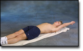
Lie on your back with the injured arm by your side. Move the arm up and over your head before slowly retuning the arm again. The arm must be outstretched the whole time.

Lift both shoulders slowly upwards and down again.

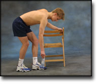
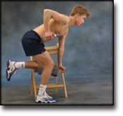
Stand bending forward and supporting a chair back with one hand. Let the other arm hang freely downwards holding a weight. Move the shoulder backwards and forwards and from side to side. It is important that the arm hangs straight down the whole time.

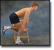
Stand slightly bent over a chair, with your weight on the good arm and the injured arm hanging loosely downwards holding a weight or a filled bottle. Lift your arm/shoulder upwards by using the shoulder blade’s muscles. Using heavier objects or weights can increase the load.

Stand at a table with the injured arm on a ball. Move the ball in all directions while applying slight pressure on the ball.

Stretching is carried out in the following way: stretch the muscle group for 3-5 seconds. Relax for 3-5 seconds. The muscle group should subsequently be stretched for 20 seconds. The muscle is allowed to be tender, but must not hurt. Relax for 20 seconds, after which the procedure can be repeated. The time consumed for stretching, coordination and strength training can be altered depending on the training opportunities available and individual requirements.

treatment-article4

SportNetDoc

Active neck muscle training in the treatment of chronic neck pain in women: a randomized controlled trial.

Ylinen J, Takala EP, Nykanen M, Hakkinen A, Malkia E, Pohjolainen T, Karppi SL, Kautiainen H, Airaksinen O. JAMA. 2003 May 21;289(19):2509-16.

CONTEXT: Active physical training is commonly recommended for patients with chronic neck pain; however, its efficacy has not been demonstrated in randomized studies. OBJECTIVE: To evaluate the efficacy of intensive isometric neck strength training and lighter endurance training of neck muscles on pain and disability in women with chronic, nonspecific neck pain. DESIGN: Examiner-blinded randomized controlled trial conducted between February 2000 and March 2002. SETTING: Participants were recruited from occupational health care systems in southern and eastern Finland. PATIENTS: A total of 180 female office workers between the ages of 25 and 53 years with chronic, nonspecific neck pain. INTERVENTIONS: Patients were randomly assigned to either 2 training groups or to a control group, with 60 patients in each group. The endurance training group performed dynamic neck exercises, which included lifting the head up from the supine and prone positions. The strength training group performed high-intensity isometric neck strengthening and stabilization exercises with an elastic band. Both training groups performed dynamic exercises for the shoulders and upper extremities with dumbbells. All groups were advised to do aerobic and stretching exercises regularly 3 times a week. MAIN OUTCOME MEASURES: Neck pain and disability were assessed by a visual analog scale, the neck and shoulder pain and disability index, and the Vernon neck disability index. Intermediate outcome measures included mood assessed by a short depression inventory and by maximal isometric neck strength and range of motion measures. RESULTS: At the 12-month follow-up visit, both neck pain and disability had decreased in both training groups compared with the control group (P<.001). Maximal isometric neck strength had improved flexion by 110%, rotation by 76%, and extension by 69% in the strength training group. The respective improvements in the endurance training group were 28%, 29%, and 16% and in the control group were 10%, 10%, and 7%. Range of motion had also improved statistically significantly in both training groups compared with the control group in rotation, but only the strength training group had statistically significant improvements in lateral flexion and in flexion and extension. CONCLUSIONS: Both strength and endurance training for 12 months were effective methods for decreasing pain and disability in women with chronic, nonspecific neck pain. Stretching and fitness training are commonly advised for patients with chronic neck pain, but stretching and aerobic exercising alone proved to be a much less effective form of training than strength training.

treatment-article3

SportNetDoc

Treatment of myofascial trigger-points with ultrasound combined with massage and exercise–a randomised controlled trial.

Gam AN, Warming S, Larsen LH, Jensen B, Hoydalsmo O, Allon I, Andersen B, Gotzsche NE, Petersen M, Mathiesen B. Pain 1998 Jul;77(1):73-9.

The effect of treatment with ultrasound, massage and exercises on myofascial trigger-points (MTrP) in the neck and shoulder was assessed in a randomised controlled trial. The outcome measures were pain at rest and on daily function (Visual Analogue Scale, VAS), analgesic usage, global preference and index of MTrP. Long-term effect for treatment and control groups was assessed after 6 months using a questionnaire. The patients were randomised to three groups. The first group was treated with ultrasound, massage and exercise (A), the second group with sham-ultrasound, massage and exercise (B), while the third group was a control group (C). The duration of the study was 6 weeks. Treatment was given twice a week from the second to the fifth week. The number and index of MTrPs were recorded at each treatment session in groups A and B but only at entry as well as end of study in group C. VAS and analgesic usage was recorded in all three groups throughout the study period. Six months after the last treatment session a questionnaire was send to the patients. A total of 67 patients were included. Nine patients dropped-out during the study, which left 58 patients that could be included in the final analysis. Twenty patients were randomised to group A, 18 to group B and 18 to group C. A significant reduction in index were found between treatment groups (A and B) and control group (C), but no difference between group A and B. VAS scores, analgesic usage or global preference showed no difference between group A, B or C. The patients in the group C were offered treatment (ultrasound, massage, exercise) after the 6 weeks treatment period. At the questionnaire after 6 month 44 (87%) of the 52 patients from all three groups who had treatment responded. Sixty-four percent answered that they had had good or some effects, 68 percent were still doing the exercise programme and 17 percent had received other forms of therapy after they had completed the study. No difference between groups given ultrasound or sham ultrasound were found. It is concluded that US give no pain reduction, but apparently massage and exercise reduces the number and intensity of MTrP. The impact of this reduction on neck and shoulder pain is weak.

treatment-article2

SportNetDoc

Rehabilitation of neck-shoulder pain in women industrial workers: a randomized trial comparing isometric shoulder endurance training with isometric shoulder strength training.

Hagberg M, Harms-Ringdahl K, Nisell R, Hjelm EW. Arch Phys Med Rehabil 2000 Aug;81(8):1051-8.

OBJECTIVES.
To study whether isometric shoulder endurance was more advantageous than isometric shoulder strength training in reducing pain and perceived exertion and to increase shoulder function through improved muscle endurance and strength.

DESIGN.
Randomized trial.

SETTING.
Three occupational health care centers. PARTICIPANTS: Women industrial workers with nonspecific neck-shoulder pain. The International Classification of Diseases, 10th Revision (ICD-10) diagnosis was “cervicobrachial syndrome” (M53.1). Thirty-eight patients completed the isometric shoulder endurance training and 31 patients completed the isometric shoulder strength training.

INTERVENTION.
Twelve weeks of training.

MAIN OUTCOME MEASURES.
Self-reported pain and rating of perceived exertion (RPE), arm motion performance test, shoulder muscle strength, shoulder muscle endurance, and shoulder functional tests, as well as follow-up after supervised training had ended.

RESULTS.
The isometric shoulder strength training resulted in an almost one-scale step decrease in RPE at work and a 5% to 15% improvement of arm motion performance compared with the endurance training. The isometric shoulder strength training more effectively improved left side shoulder abduction strength (p < .026), but no major differences were found for the other strength measurements. The isometric shoulder endurance training was not more successful than the strength training in the endurance test (p .51 to .81).

CONCLUSIONS.
Physical training programs for neck-shoulder pain may include isometric shoulder muscular strength exercise in addition to isometric shoulder endurance training, rather than endurance training only.

treatment-article1

SportNetDoc

Perceived pain before and after three exercise programs–a controlled clinical trial of women with work-related trapeziusP myalgia.

Waling K, Sundelin G, Ahlgren C, Jarvholm B. Pain 2000 Mar;85(1-2):201-7.

The effect of exercise on neck-shoulder pain was studied in 103 women with work-related trapezius myalgia randomized into three exercise groups and a control group. One group trained strength, the second muscular endurance and the third co-ordination. The exercise groups met three times weekly for 10 weeks. Pain assessment was made on three visual analogue scales, indicating pain at present, pain in general and pain at worst. Pain thresholds were measured in the trapezius muscle with a pressure algometer. A pain drawing was completed. The rated pain decreased significantly (P<0.05) on the VAS describing pain at worst in the strength and endurance groups. Pressure sensitivity decreased significantly (P<0.05) in four triggerpoints in the exercise groups. No changes were seen in the extent of painful body area in any group. Comparison of exercisers (n=82) and controls (n=21) showed significantly larger pain reductions on VAS pain at present and VAS pain at worst among exercisers. All three exercise programs showed similar decreases of pain which indicates that the type of exercise is of less importance to achieve pain reduction.

treatment-article

SportNetDoc

Do Colles’ fracture patients benefit from routine referral to physiotherapy following cast removal?

Watt CF, Taylor NF, Baskus K. Arch Orthop Trauma Surg 2000;120(7-8):413-5.

Colies’ fracture patients who received physiotherapy immediately following cast removal were compared with patients who received no active therapy following cast removal in a prospective randomised study. Patients who attended physiotherapy achieved significantly greater increases in wrist extension and grip strength after 6 weeks compared to patients who received no active therapy.

complication-article

SportNetDoc

Fractures of the scaphoid.

Mansat M. Ann Chir Main 1982;1(4):361-74.

Scaphoid fractures are the most common carpal injury. The diagnosis is often difficult and the treatment often controversial. Late complications are frequent and include nonunion, avascular necrosis and arthrosis. Carpal pathomechanics, the scaphoid’s blood supply and fracture healing are basic concepts leading to a better understanding of the injury. This paper discusses the scaphoid in relation to the pathomechanics of the wrist, the relationship of the blood supply of the scaphoid to the injury and the diagnosis, classification and treatment of these fractures.

treatment-article

SportNetDoc

Scaphoid fracture. Review of diagnostic tests and treatment.

Schubert HE. Can Fam Physician 2000 Sep;46:1825-32.

OBJECTIVE.
To help make diagnosis and treatment of scaphoid fracture more precise by review of published evidence.

QUALITY OF EVIDENCE.
MEDLINE was searched using the terms “scaphoid,” “carpal navicular,” “fracture,” “computed tomography,” “bone scan,” and “scintigraphy.” Most papers were case-series observational reports. Papers were cited if the case series was large or if there was a high degree of agreement among several observers. The main recommendation for change in treatment of scaphoid fracture is based on two randomized clinical trials involving more than 1000 patients with proven scaphoid fracture.

MAIN MESSAGE.
Fracture of the scaphoid requires a specific mechanism of injury. “Snuffbox” tenderness is not specific for scaphoid fracture and is not the most useful physical finding; other physical findings provide more specific evidence for or against scaphoid fracture. Physical examination remains the basis of initial treatment and should be thorough and meticulous. X-ray films must be of high quality and should be examined carefully for bone and soft tissue signs of fracture. A Colles’-type short arm cast is adequate for treating common undisplaced scaphoid waist fractures; the thumb need not be immobilized. For suspected scaphoid fractures, without radiologic evidence of fracture, treating symptoms is likely sufficient.

CONCLUSION.
Evidence found in the literature can be used to improve diagnostic accuracy for scaphoid fractures, to optimize treatment for these injuries, and to reduce unnecessary immobilization and disability for patients.

treatment-article

SportNetDoc

Metacarpal and phalangeal fractures in athletes.

Capo JT, Hastings H 2nd. Clin Sports Med 1998 Jul;17(3):491-511.

The high demands placed on the upper extremity in sporting activities subject the competitive athlete to common injuries of the hand. Treatment options are based on the fracture configuration, associated extremity injuries, and status of the surrounding soft tissue. Metacarpal and phalangeal fractures may usually be treated by closed, nonoperative methods, and most athletes may quickly return to play with a protective orthosis. Supplemental methods of fixation, such as percutaneous pins and tension-band wires, may be used for unstable fractures. When required, open reduction and internal fixation can provide optimum stability to the fracture, which allows immediate range-of-motion and early return to play.

complication-article

SportNetDoc

Hyperextension injury to the PIP joint or to the MP joint of the thumb–a clinical study.

Jespersen B, Nielsen NS, Bonnevie BE, Boeckstyns ME. Scand J Plast Reconstr Surg Hand Surg 1998 Sep;32(3):317-21.

We present a prospective study of the diagnosis and clinical course of 60 patients with 57 pure hyperextension injuries to the proximal interphalangeal (PIP) joint of the long fingers (fingers 2-5) and seven injuries to the metacarpophalangeal (MP) joint of the thumb. Thirty four of the injuries (57%) were related to ball sports, and the ulnar fingers of the non-dominant hand were usually affected. There were 24 avulsion fractures at the site of the insertion of the volar plate on to the middle phalanx. Twelve (20%) initially presented with hyperextension instability, and this was usually associated with an avulsion fracture. Thirty four of the patients (57%) had symptoms for less than one month, while 10 (17%) complained of symptoms six months after the injury. Severe complications such as daily pain and stiffness were encountered in three cases. The triad sign (pain on extreme flexion and extension) was of no use as a diagnostic or prognostic factor, nor did the radiographic stress-view help to identify acute instability of the joint.