Alle indlæg af urtehave_com

KONDITION

STEP3

Training ladder for:
METATARSUS LIGAMENT RUPTURE
(RUPTURA TRAUMATICA LIGAMENTI PEDIS)

STEP 3

KONDITION
Unlimited: Cycling. Swimming. Running straight ahead (without directional change).

UDSPÆNDING
(10 min)

Stand with the injured leg stretched backwards with the toes facing front. Slowly bend the knee so that the calf muscles become increasingly stretched. Hold the position for 20 seconds and relax for 20 seconds before repeating.

Stand with the injured leg outstretched with the ankle joint bent up against the wall. Press your abdomen against the wall so that the calf muscles become increasingly stretched. Hold the position for 20 seconds and relax for 20 seconds before repeating.

Stand on the good leg while gaining support on the wall with the hand on the same side. Bend the knee of the injured leg and draw the heel towards the buttocks. Take hold of the big toe side of the foot and draw slowly upwards so that the ankle joint is stretched to the maximum and drawn slightly outwards so that increased stretching is experienced on the inside of the shin bone. Hold the position for 20 seconds and relax for 20 seconds before repeating.

Stand on the good leg while gaining support from the wall with the opposite hand. Bend the knee of the injured leg and draw the heel towards the buttocks. Take hold of the little toe side of the foot and draw slowly upwards so that the ankle joint is stretched to the maximum and drawn over the good leg so that increased stretching is experienced on the outer side of the shin bone. Hold the position for 20 seconds and relax for 20 seconds before repeating.

KOORDINATION
(10 min)

Stand on the leg to be trained. Take-off and land on the same leg.

Seesaw. Balance on two legs, possibly using a hand as support against the wall, balancing subsequently on one leg without support. Look straight ahead and keep knees bent.

Stand on the injured leg with your upper body bent forwards at 90 degrees. Lift the good leg in a straight line behind you. When you feel comfortable with the exercise, it can be made more difficult by closing your eyes.

STYRKE
(40 min)

Sit on the floor. Tip the foot up and down with elastic under the forefoot so that the elastic becomes taut.

Sit on the floor. Tip the foot up and down with elastic on top of the forefoot so that the elastic becomes taut.

Sit on the floor. Tip the foot from side to side with elastic on the outer side of the foot, without moving the knee.

Sit on the floor. Tip the foot from side to side with elastic on the inner side of the foot, without moving the knee.

Stand behind a chair. Rise slowly up on tiptoe and go down again.

Up and down from the stool with load. Tie elastic around the hip and go up on the stool in a slow movement. The elastic should be fastened to the wall.

Go up and down from the stool. Go up with alternating right and left legs.

Stand on the healthy leg with elastic fixed around the hip. The elastic should be fixed to the wall or a wall bar. Take-off on the healthy leg and land on the leg to be trained and keep your balance. Remember that the elastic should be positioned so that it gives resistance at the moment of take-off. Change legs.

Sit with your bare toes on a tea towel and curl the tea towel together using your toes.

Stand with both legs on the stool with elastic around the hip. Take-off and land with feet together.

Stretching is carried out in the following way: stretch the muscle group for 3-5 seconds. Relax for 3-5 seconds. The muscle group should subsequently be stretched for 20 seconds. The muscle is allowed to be tender, but must not hurt. Relax for 20 seconds, after which the procedure can be repeated. The time consumed for stretching, coordination and strength training can be altered depending on the training opportunities available and individual requirements.

KONDITION

STEP2

Training ladder for:
METATARSUS LIGAMENT RUPTURE
(RUPTURA TRAUMATICA LIGAMENTI PEDIS)

STEP 2

KONDITION
Unlimited: Cycling. Swimming. Light running straight ahead (without directional change) on a smooth surface.

UDSPÆNDING
(10 min)

Stand with the injured leg stretched backwards with the toes facing front. Slowly bend the knee so that the calf muscles become increasingly stretched. Hold the position for 20 seconds and relax for 20 seconds before repeating.

Stand with the injured leg outstretched with the ankle joint bent up against the wall. Press your abdomen against the wall so that the calf muscles become increasingly stretched. Hold the position for 20 seconds and relax for 20 seconds before repeating.

Stand on the good leg while gaining support on the wall with the hand on the same side. Bend the knee of the injured leg and draw the heel towards the buttocks. Take hold of the big toe side of the foot and draw slowly upwards so that the ankle joint is stretched to the maximum and drawn slightly outwards so that increased stretching is experienced on the inside of the shin bone. Hold the position for 20 seconds and relax for 20 seconds before repeating.

Stand on the good leg while gaining support from the wall with the opposite hand. Bend the knee of the injured leg and draw the heel towards the buttocks. Take hold of the little toe side of the foot and draw slowly upwards so that the ankle joint is stretched to the maximum and drawn over the good leg so that increased stretching is experienced on the outer side of the shin bone. Hold the position for 20 seconds and relax for 20 seconds before repeating.

KOORDINATION
(10 min)

Stand on one leg. Play the ball up against the wall.

Seesaw. Balance on two legs, possibly using a hand as support against the wall, balancing subsequently on one leg without support. Look straight ahead and keep knees bent.

STYRKE
(40 min)

Sit on the floor. Tip the foot up and down with elastic under the forefoot so that the elastic becomes taut.

Sit on the floor. Tip the foot up and down with elastic on top of the forefoot so that the elastic becomes taut.

Sit on the floor. Tip the foot from side to side with elastic on the outer side of the foot, without moving the knee.

Sit on the floor. Tip the foot from side to side with elastic on the inner side of the foot, without moving the knee.

Stand behind a chair. Rise slowly up on tiptoe and go down again.

Up and down from the stool with load. Tie elastic around the hip and go up on the stool in a slow movement. The elastic should be fastened to the wall.

Go up and down from the stool. Go up with alternating right and left legs.

Sit with your bare toes on a tea towel and curl the tea towel together using your toes.

Stand with both legs on the stool with elastic around the hip. Take-off and land with feet together.

Stretching is carried out in the following way: stretch the muscle group for 3-5 seconds. Relax for 3-5 seconds. The muscle group should subsequently be stretched for 20 seconds. The muscle is allowed to be tender, but must not hurt. Relax for 20 seconds, after which the procedure can be repeated. The time consumed for stretching, coordination and strength training can be altered depending on the training opportunities available and individual requirements.

KONDITION

STEP1

Training ladder for:
METATARSUS LIGAMENT RUPTURE
(RUPTURA TRAUMATICA LIGAMENTI PEDIS)

STEP 1

The indications of time after stretching, coordination training and strength training show the division of time for the respective type of training when training for a period of one hour. The time indications are therefore not a definition of the daily training needs, as the daily training is determined on an individual basis.

KONDITION
Unlimited: Cycling. Swimming. Running in deep water.

UDSPÆNDING
(10 min)

Stand with the injured leg stretched backwards with the toes facing front. Slowly bend the knee so that the calf muscles become increasingly stretched. Hold the position for 20 seconds and relax for 20 seconds before repeating.

Stand with the injured leg outstretched with the ankle joint bent up against the wall. Press your abdomen against the wall so that the calf muscles become increasingly stretched. Hold the position for 20 seconds and relax for 20 seconds before repeating.

Stand on the good leg while gaining support on the wall with the hand on the same side. Bend the knee of the injured leg and draw the heel towards the buttocks. Take hold of the big toe side of the foot and draw slowly upwards so that the ankle joint is stretched to the maximum and drawn slightly outwards so that increased stretching is experienced on the inside of the shin bone. Hold the position for 20 seconds and relax for 20 seconds before repeating.

Stand on the good leg while gaining support from the wall with the opposite hand. Bend the knee of the injured leg and draw the heel towards the buttocks. Take hold of the little toe side of the foot and draw slowly upwards so that the ankle joint is stretched to the maximum and drawn over the good leg so that increased stretching is experienced on the outer side of the shin bone. Hold the position for 20 seconds and relax for 20 seconds before repeating.

KOORDINATION
(10 min)

Stand on one leg on the floor or a mattress. Look straight ahead and keep the knee slightly bent.

STYRKE
(40 min)

Sit on the floor. Tip the foot up and down with elastic under the forefoot so that the elastic becomes taut.

Sit on the floor. Tip the foot up and down with elastic on top of the forefoot so that the elastic becomes taut.

Sit on the floor. Tip the foot from side to side with elastic on the outer side of the foot, without moving the knee.

Sit on the floor. Tip the foot from side to side with elastic on the inner side of the foot, without moving the knee.

Stand on a soft surface. Rise slowly up on tiptoe and go down again.

Go up and down from the stool. Go up with alternating right and left legs.

Sit with a ball under the foot. Roll the ball backwards and forwards and from side to side.

Stand on both legs. Tip the toes on the leg to be trained upwards and down again, whilst having the heel firmly on the floor during the exercise.

Sit with your bare toes on a tea towel and curl the tea towel together using your toes.

Sit on a chair. Keep the heel firmly on the ground and tip the toes up.

Stretching is carried out in the following way: stretch the muscle group for 3-5 seconds. Relax for 3-5 seconds. The muscle group should subsequently be stretched for 20 seconds. The muscle is allowed to be tender, but must not hurt. Relax for 20 seconds, after which the procedure can be repeated. The time consumed for stretching, coordination and strength training can be altered depending on the training opportunities available and individual requirements.

tape-instruction

Tapening


Type: METATARSUS:

Objective: Support, and therefore relieve, the metatarsi bones.

Application: A tape “anchor” is applied around the forefoot behind the toe pad (A). A new strip is applied in the same manner half overlapping the first and a little behind (B). If it is a large foot a third strip can be applied a further half tape’s width behind.

paracetamol-a1

SportNetDoc

Relative risk of upper gastrointestinal complications among users of acetaminophen and nonsteroidal anti-inflammatory drugs.

Garcia Rodriguez LA, Hernandez-Diaz S. Epidemiology 2001 Sep;12(5):570-6

Nonsteroidal anti-inflammatory drugs (NSAIDs) have been associated with an increase in upper gastrointestinal complications. There is no agreement, however, on whether all conventional NSAIDs have a similar relative risk (RR), and epidemiologic data are limited on acetaminophen. We studied the association between these medications and the risk of upper gastrointestinal bleed/perforation in a population-based cohort of 958,397 persons in the United Kingdom between 1993 and 1998. Our nested case-control analysis included 2,105 cases and 11,500 controls. RR estimates were adjusted for several factors known to be associated with upper gastrointestinal bleed/perforation. Compared with non-users, users of acetaminophen at doses less than 2 gm did not have an increased risk of upper gastrointestinal complications. The adjusted RR for acetaminophen at doses greater than 2 gm was 3.6 [95% confidence interval (95% CI) = 2.6-5.1].
The corresponding RRs for low/medium and high doses of NSAIDs were 2.4 (95% CI = 1.9-3.1) and 4.9 (95% CI = 4.1-5.8). The RR was 3.1 (95% CI = 2.5, 3.8) for short plasma half-life, 4.5 (95% CI = 3.5-5.9) for long half-life, and 5.4 (95% CI = 4.0-7.1) for slow-release formulations of NSAIDs.
After adjusting for daily dose, the differences in RR between individual NSAIDs tended to diminish except for apazone. Users of H2 receptor antagonists, omeprazole, and misoprostol had RRs of 1.4 (95% CI = 1.2-1.8), 0.6 (95% CI = 0.4-0.9), and 0.6 (95% CI = 0.4-1.0), respectively. Among NSAID users, use of nitrates was associated with an RR of 0.6 (95% CI = 0.4-1).

tape-instruction

Tapening


Type: BIG TOE:

Objective: Support, and therefore relieve, the metarsophalangeal joint of the big toe.

Application: A tape “anchor” is applied around the forefoot behind the toe pad (A). 2-3 strips are applied from the anchor on the back of the foot around the big toe, and back to the anchor on the back of the foot (B).

paracetamol

Paracetalmol

SIMPLE ANALGESIC.

Indication. Analgesic drugs (painkillers) can be used to a limited extent to reduce the pain with minor injuries where there are no risks of aggravating the injury through continued sports activity (i.e. bleeding under the nail, skin abrasions etc.). All forms of painkillers can naturally be taken if the sports activity ceases. Paracetamol is recommended due to the very modest side effects associated with its use.

Mechanism of action. Paracetamol provides both painkilling (analgesic) and temperature lowering (antipyretic) effects. The mechanism is partly unknown. 90% is absorbed from the intestines after ingestion of a tablet, with maximum concentration of paracetamol being achieved after ½-1 hour after ingestion. The duration of the effect is 4-6 hours. It should be noted that only approx. 60% is absorbed from suppositories.

Side effects. Paracetamol in normal doses has by and large no side effects, as opposed to weak morphine type drugs containing acetyl salicylic acid. Long term treatment with maximum dosage appears, however, to increase the risk of ulcers, especially if the treatment is combined with rheumatic pills (NSAID), (article).

Contraindications. Painkillers should never be used to enable the athlete to continue an activity which bears a risk of aggravating the injury. Paracetamol and other painkilling drugs with antipyretic effects must never be used to lower the body temperature of an athlete before starting sports activity. Serious virus infections can invade the (cardiac) muscle of the heart and cause myocarditis. The risk of myocarditis is increased under great physical exertion during virus infections, and some cases have been reported of fatalities amongst young, otherwise healthy athletes, under these circumstances. Increased body temperature indicates an infection, and all athletes should stop sports activity until the body temperature has returned to normal.
Dose. 1 gram paracetamol 3-4 times daily.

Conclusion. Paracetamol can be recommended when needed as painkillers for conditions where pain is experienced without any suspicions of inflammation.

Cartilage damage in the joint

CARTILAGE DAMAGE IN THE FOOT

Diagnosis: CARTILAGE DAMAGE IN THE FOOT


Anatomy:
The surfaces of the joints are lined with a cartilage covering of a few millimetre’s thickness which serves to reduce the load or strain on the joint surfaces.

  1. Phalanx media
  2. Tuberositas ossis metatarsalis V
  3. Os cuboideum
  4. Calcaneus
  5. Talus
  6. Os naviculare
  7. Os cuneiforme laterale
  8. Os cuneiforme intermedium
  9. Os cuneiforme mediale
  10. Os metatarsalei
  11. Os sesamoideum
  12. Phalanx proximalis
  13. Phalanx distalis

THE FOOT FROM ABOVE

Cause: Localised cartilage injuries in the joint surfaces can occur after a vigorous twisting of the joint, where the joint surfaces impact on each other and cause cartilage damage. In some cases a piece of cartilage can be shed which can wander in the joint (joint mouse) and become jammed.

Symptoms: Pain in the joint when under load or strain. Occasional inflammation of the synovial membrane which causes concentration of fluid in the joint.

Examination: Normal medical examination is often not sufficient. To make the diagnosis correctly it is therefore necessary to perform an arthroscopic examination or an MR-scan.

Treatment: Treatment comprises relief from the painful activities until the pain is no longer experienced, after which gradual training can be commenced. There is no treatment that can restore the damaged cartilage, which has itself poor restorative ability. Different procedures to enhance the healing can be attempted using arthroscopic examination, however, the results are generally unsatisfactory (article-1) (article-2). Results from experimental cartilage transplants are still not successful enough to warrant introduction as a routine treatment in the near future. Joint mouse which provokes the symptoms must be surgically removed.

Rehabilitation: Rehabilitation is completely dependent upon the type of cartilage damage (size and position in the joint) and treatment (conservative or surgical).
Also read rehabilitation, general.

Complications: Greater cartilage injuries which are positioned on the weight-bearing parts of the joint are some of the most serious sports injuries, and often results in an end to the sporting career.

Special: As there is a risk that the injury can be permanent, all cases should be reported to your insurance company.

treatment-article1

SportNetDoc

Interventions for preventing and treating stress fractures and stress reactions of bone of the lower limbs in young adults.

Gillespie WJ, Grant I. Cochrane Database Syst Rev 2000;(2):CD000450.

BACKGROUND.
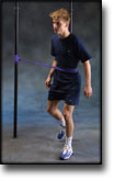
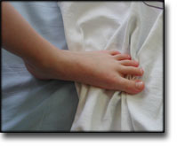
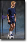
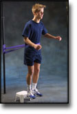
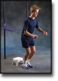
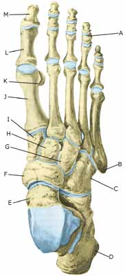
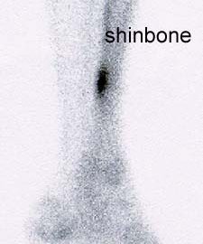
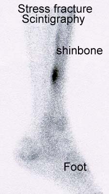
Stress reaction in bone, which may proceed to a fracture, is a significant problem in military recruits and in athletes, particularly long distance runners.

OBJECTIVES.
To evaluate the evidence from controlled trials of treatments and programmes for prevention or management of lower limb stress fractures and stress reactions of bone in active young adults.

SEARCH STRATEGY.
We searched the Cochrane Musculoskeletal Injuries Group Trials Register, The Cochrane Library, MEDLINE, EMBASE, Current Contents, Dissertation Abstracts, Index to UK Theses and the bibliographies of identified articles. Date of last search: December 1997.

SELECTION CRITERIA.
Any randomised or quasi-randomised trial evaluating a programme or treatment to prevent or treat lower limb stress reactions of bone or stress fractures in active young adults.

DATA COLLECTION AND ANALYSIS.
Searching, a decision on inclusion or exclusion, methodological assessment, and data extraction were carried out according to a predetermined protocol included in the body of the review. Analysis using Review Manager software allowed pooling of data and calculation of Peto odds ratios and absolute risk reductions, each with 95% confidence intervals.

MAIN RESULTS.
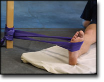
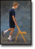
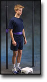
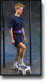
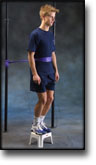
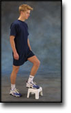
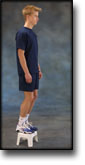
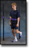
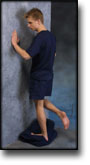
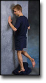
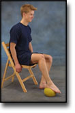
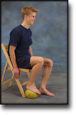
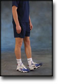
The use of “shock absorbing” insoles, evaluated in four trials, appears to reduce the incidence of stress fractures and stress reactions of bone (Peto odds ratio 0.47, 95% confidence interval 0. 30 to 0.76). Incomplete data from one trial indicated that reduction of running and jumping intensity may also be effective. The use of pneumatic braces in the rehabilitation of tibial stress fractures significantly reduces the time to recommencing training (weighted mean difference -42.6 days, 95% confidence interval -55.8 to -29.4 days).

REVIEWER’S CONCLUSIONS.
The use of shock absorbing insoles in footwear reduces the incidence of stress fractures in athletes and military personnel. Rehabilitation after tibial stress fracture is aided by the use of pneumatic bracing.