|
Relative risk of upper gastrointestinal complications among users of acetaminophen and nonsteroidal anti-inflammatory drugs. |
Kategoriarkiv: Degenerative arthritis
tape-instruction
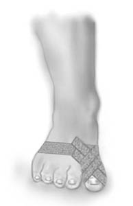
Objective: Support, and therefore relieve, the metarsophalangeal joint of the big toe. |
|||
 |
|||
| Application: A tape “anchor” is applied around the forefoot behind the toe pad (A). 2-3 strips are applied from the anchor on the back of the foot around the big toe, and back to the anchor on the back of the foot (B). |
Cartilage damage in the joint
|
||
|
||
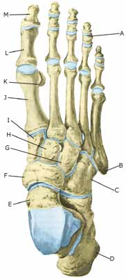
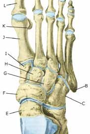
| Cause: Localised cartilage injuries in the joint surfaces can occur after a vigorous twisting of the joint, where the joint surfaces impact on each other and cause cartilage damage. In some cases a piece of cartilage can be shed which can wander in the joint (joint mouse) and become jammed.
Symptoms: Pain in the joint when under load or strain. Occasional inflammation of the synovial membrane which causes concentration of fluid in the joint. Examination: Normal medical examination is often not sufficient. To make the diagnosis correctly it is therefore necessary to perform an arthroscopic examination or an MR-scan. Treatment: Treatment comprises relief from the painful activities until the pain is no longer experienced, after which gradual training can be commenced. There is no treatment that can restore the damaged cartilage, which has itself poor restorative ability. Different procedures to enhance the healing can be attempted using arthroscopic examination, however, the results are generally unsatisfactory (article-1) (article-2). Results from experimental cartilage transplants are still not successful enough to warrant introduction as a routine treatment in the near future. Joint mouse which provokes the symptoms must be surgically removed. Rehabilitation: Rehabilitation is completely dependent upon the type of cartilage damage (size and position in the joint) and treatment (conservative or surgical). Complications: Greater cartilage injuries which are positioned on the weight-bearing parts of the joint are some of the most serious sports injuries, and often results in an end to the sporting career. Special: As there is a risk that the injury can be permanent, all cases should be reported to your insurance company. |