Kategoriarkiv: Arm, lower

Inflammation of the bursa

INFLAMMATION OF THE BURSA

Diagnosis: INFLAMMATION OF THE BURSA
(Bursitis)


Anatomy:
On the forearm there are numerous bursas reducing the pressure on muscles and tendons, where these lie close to the bone.

Cause: In case of repeated loads or blows the bursas can become inflamed, produce fluid, swell and become painful. One of the most frequent bursitis forms on the forearm, is inflammation of the bursa located between the biceps tendon and the fastening on the radius (tuberositas radii) (article).

Symptoms: Pain upon applying pressure on the bursa, which sometimes (but far from always) may feel swollen. Aggravated upon activation of the muscle located immediately above the bursa.

Acute treatment: Click here.

Examination: In slight cases with only minimal tenderness, medical examination is not necessarily required. In cases of more pronounced pain or lack of progress, a medical examination should be carried out to ensure a correct diagnosis and treatment. The diagnosis is usually made on the basis of a normal medical examination, however, if there is any doubt surrounding the diagnosis, it can easily and quickly be confirmed under an ultrasound scan.

Treatment: The treatment primarily consists of relief, and removal of the provoking cause (if identified). The treatment can be supplemented with rheumatic medicine (NSAID) or the injection of corticosteroid in the bursa preceded by draining of the bursa, which can advantageously be done under ultrasound guidance.

Rehabilitation: The treatment is completely dependant on which bursa is inflamed, but sports activity can usually be cautiously resumed once pain has decreased, particularly if it has been possible to remove the provoking cause.
Also read rehabilitation, general.

Complications: If progress is not smooth, it should be considered if the diagnosis is correct or whether complications have arisen. In rare cases the bursa can become infected with bacteria. This is a serious condition where the bursa becomes red, warm and increasingly swollen and tender, and requires immediate medical examination and treatment.

If there is no progress with relief, medical treatment (rheumatic medicine (NSAID) and the ultrasound guided injection of corticosteroid), surgical removal of the bursa may be attempted.

Chronic compartment syndrome

CHRONIC COMPARTMENT SYNDROME

Diagnosis: CHRONIC COMPARTMENT SYNDROME


Anatomy:
The forearm muscles are divided into three groups (muscle compartments) of powerful, partially unyielding, muscle membranes (fascias); a front, an outer and a rear muscle compartment. Each muscle group has its own blood and nerve supply.

Cause: The muscles can increase so quickly following intensive training of the forearm that the muscle membranes surrounding the muscles cannot keep up, causing the pressure in the muscle compartment to increase. The pressure can in some cases increase so greatly that impingement of blood vessels and nerves can occur. In other cases, chronic muscle compartment syndrome can arise due to development of scar tissue in the muscle (following previous muscle ruptures).

Symptoms: With chronic compartment syndrome there is slowly insetting pain in the muscles after a few minutes activity. There is a sensation that the muscle is “tightened” and becomes hard, which is accompanied by discomfort. If the activity is stopped the discomfort diminishes, but returns after a short period of resuming the sports activity once again. There is often sensory disturbance in the fingers.

Examination: The diagnosis is made on the basis of the characteristic history and possibly with a pressure measurement in the muscle compartment.

Treatment: With chronic muscle compartment syndrome the treatment primarily comprises relief and slowly increasing training intensity and possibly rheumatic medicine (NSAID). If there is scar tissue in the muscle, ultrasound guided injection of corticosteroid around the scar tissue formation can be attempted. In cases where there is a lack of progress a surgical splitting of the muscle membranes can be performed, which is usually a minor procedure with good results (article).

Rehabilitation: INSTRUCTION

Rehabilitation: Rehabilitation is dependant upon which muscle group has been affected. Once the pain has diminished, the sports activity can generally be slowly resumed according to the principles mentioned under rehabilitation, general.

Complications: In cases of lack of progress with relief and slow rehabilitation, an ultrasound scan should be performed before possible surgery to rule out complications from an earlier muscle rupture.

Acute compartment syndrome

ACUTE COMPARTMENT SYNDROME

Diagnosis: ACUTE COMPARTMENT SYNDROME


Anatomy:
The forearm muscles are divided into three groups (muscle compartments) of powerful, partially unyielding, muscle membranes (fascias); a front, an outer and a rear muscle compartment. Each muscle group has its own blood and nerve supply.

Cause: The pressure in a muscle compartment can rise so fast (due to bleeding or fluid extraction) that the muscle membranes cannot keep up. The pressure in the muscle compartment can therefore increase so greatly that impingement of blood vessels and nerves can occur (article).

Symptoms: With the acute muscle compartment syndrome there is increasing pain, which is often more powerful than expected from the primary evaluation of the extent of the injury. At the same time sensory disturbances can occur in the fingers.

Acute treatment: Click here.

Examination: The diagnosis is made on the basis of the characteristic history, and by a pressure measurement in the muscle compartment.

Treatment: With acute muscle compartment syndrome the treatment in severe cases comprises acute splitting of the muscle membrane. It is imperative for the continuing function of the muscle that this operation be acute, which is, of course, only possible if the athlete seeks acute medical attention (article-1) (article-2).

Rehabilitation: The rehabilitation is completely dependant on which muscle groups are affected, the provoking cause (blow to the muscle, muscle rupture or over-training) and which treatment that has been performed (relief, surgical splitting). Once pain has decreased, the sports activity can generally be slowly resumed according to the principles as mentioned under rehabilitation, general.

Complications: Muscles and nerves can suffer permanent damage if the treatment is not started as soon as possible.

Special: Since there is a risk of permanent disability, the injury should be reported to your insurance company.

Bone fracture

BONE FRACTURE

Diagnosis: BONE FRACTURE
(Fractura antebrachii)


Anatomy:
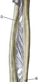
The forearm bones consist of the ulna and the radius.

  1. Elbow
  2. Ulna
  3. Wrist
  4. Radius

FOREARM BONES FROM THE FRONT

Cause: A bone fracture can occur in cases of a direct blow or fall on the arm, and can occur anywhere on the bone. It is often seen in children that the bone merely “bends” (green-stick fracture).

Symptoms: Sudden pain and pain induced constriction of movement of the arm after a fall or blow. An angling of the forearm can occasionally be seen. In rare case, acute compartment syndrome can develop.

Acute treatment: Click here.

Examination: Sudden, powerful pains in the arm with constriction of movement after a fall, should always lead to acute medical examination. The fracture is usually visible on x-rays, and on the basis of the type of fracture, the correct treatment can be determined.

Treatment: In cases of considerable dislocation or angling of the bones, the fracture will be reset under an anaesthetic, followed by bandaging for a few weeks. In certain types of fractures, an operative fixation can be necessary.

Rehabilitation: When pain has decreased fitness training in the form of cycling may be started along with rehabilitation as specified under rehabilitation, general. The rehabilitation period is completely dependent upon the type of fracture and the treatment administered.

Bandage: Special plastic bandages can be made for use following a fracture of the forearm when sports are resumed.

Complications: In the vast majority of cases the fracture heals without complications, although in some cases a poor healing occurs which can affect the blood vessel and the nerve supply to the arm, resulting in chronic compartment syndrome. If satisfactory progress is not achieved, you should therefore consult your doctor.