Kategoriarkiv: Thigh, front

Stress fracture

STRESS FRACTURE

Diagnosis: STRESS FRACTURE
(Stress fracture)


Anatomy:
The femur is the only bone in the thigh. Innumerable muscles are attached to the bone.


  1. Caput femoris
  2. Collum femoris
  3. Trochanter minor
  4. Trochanter major

THIGH BONE FROM THE FRONT

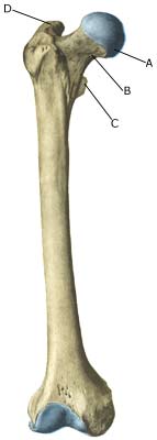
Cause: Repeated loads, particularly when walking or running, can cause such great stress that cracks (stress fractures) appear in the shaft of the femur (article).

Symptoms: Pain and tired sensation in the thigh. The pain is aggravated upon applying pressure (direct and indirect tenderness) and applying load (walking, running).

Examination: X-ray. Since many stress fractures are not visible early in the course, x-ray examination can be repeated after a few weeks, if stress fractures are still suspected. Scintigraphy, CT, MRI and ultrasound scans can often diagnose stress fractures far earlier than x-rays (Ultrasonic image). The frequency of stress fractures in the femur is probably more often than presumed (article). It is crucial for the result of the treatment that the diagnosis is made as early as possible (article).

Treatment: The treatment primarily comprises relief. Only in special cases is surgery necessary (article).

Rehabilitation: The rehabilitation is completely dependant on the type of fracture and the treatment (relief or surgical). A rehabilitation period of 2-4 months must be expected before maximum participation in sports activity can be resumed (article).

Complications: If progress is not smooth, you should be medically re-evaluated to ensure that the fracture is healing according to plan. In some cases a false joint is formed (pseudoarthrosis), which requires surgical treatment.

Chronic compartment syndrome

CHRONIC COMPARTMENT SYNDROME

Diagnosis: CHRONIC COMPARTMENT SYNDROME
(Chronic compertment syndrome)


Anatomy:
The thigh muscles are divided into three groups (muscle compartments) of powerful, partially unyielding, muscle membranes (fascias); a front, an inner and a rear muscle compartment (Drawing).

Cause: The muscles can increase so quickly following intensive training that the muscle membranes surrounding the muscles cannot keep up, causing the pressure in the muscle compartment to increase. The pressure can in some cases increase so greatly that impingement of blood vessels and nerves can occur. In other cases, chronic muscle compartment syndrome can arise as a complication to earlier muscle ruptures.

Symptoms: With chronic compartment syndrome there is slowly insetting pain in the anterior muscles after a few minutes activity. There is a sensation that the muscle is “tightened” and becomes hard, which is accompanied by discomfort. If the activity is stopped the discomfort diminishes, but returns after a short period of resuming the sports activity once again.

Examination: The diagnosis is made on the basis of the characteristic history and possibly with a pressure measurement in the muscle compartment (article).

Treatment: With chronic muscle compartment syndrome the treatment primarily comprises relief and slowly increasing training intensity, rheumatic medicine (NSAID), and massage (article). If there is scar tissue in the muscle, ultrasound guided injection of corticosteroid around the scar tissue formation can be attempted. In cases where there is a lack of progress a surgical splitting of the muscle membranes can be performed, which is usually a minor procedure with good results (article-1) (article-2).

Once the pain has diminished, the sports activity can generally be slowly resumed according to the principles mentioned under rehabilitation, general.

Complications: In cases of lack of progress with relief and slow rehabilitation, an ultrasound scan should be performed before possible surgery to rule out scar tissue formation in the muscles from previous muscle ruptures. Ultrasound guided injection of corticosteroid can be attempted in the area surrounding the scar tissue if scar tissue is in evidence in the muscles.

Acute compartment syndrome

ACUTE COMPARTMENT SYNDROME

Diagnosis: ACUTE COMPARTMENT SYNDROME
(Acute compartment syndrome)


Anatomy:
The thigh muscles are divided into three groups (muscle compartments) of powerful, partially unyielding, muscle membranes (fascias); a front, an inner and a rear muscle compartment (Drawing).

Cause: The pressure in a muscle compartment can rise so fast (due to bleeding or fluid extraction) that the muscle membranes cannot keep up. The pressure in the muscle compartment can therefore increase so greatly that impingement of blood vessels and nerves can occur.

Symptoms: With the acute muscle compartment syndrome there is increasing pain, which is often more powerful than expected from the primary evaluation of the extent of the injury. At the same time sensory disturbances can occur in the lower leg and feet.

Acute treatment: Click here.

Examination: The diagnosis is made on the basis of the characteristic history, increased circumference of the thigh, tight and hard anterior thigh muscle and by a pressure measurement in the muscle compartment (article).

Treatment: With acute muscle compartment syndrome the treatment in severe cases comprises acute splitting of the muscle membrane. It is imperative for the continuing function of the muscle that this operation be acute, which is, of course, only possible if the athlete seeks acute medical attention (article-1) (article-2). In mild cases, surgery can be omitted under close hospital observation (article).

Rehabilitation: The rehabilitation is completely dependant on which muscle groups are affected, the provoking cause (blow to the muscle, muscle rupture or over-training) and which treatment that has been performed (relief, surgical splitting). Once pain has decreased, the sports activity can generally be slowly resumed according to the principles as mentioned under rehabilitation, general.

Complications: Muscles and nerves can suffer permanent damage if the treatment is not started as soon as possible.

Special: Since there is a chance of permanent disability, the injury should be reported to your insurance company.

Muscular bleeding in the anterior thigh

MUSKELBLØDNING I FORLÅR

Diagnosis: MUSCULAR BLEEDING IN THE ANTERIOR THIGH
(Haematoma musculi)


Anatomy:
The thigh muscles are dealt up in three muscle groups (muscle compartments) of powerful, partially unyielding, muscle membranes (fascias): a front, an inner and a rear muscle compartment. The anterior thigh muscle consists of four muscles (M vastus lateralis, M vastus medialis, M rectus femoris and the deep lying M vastus intermedius).


  1. Spina iliaca anterior superior
  2. M. iliopsoas
  3. Lig. inguinale
  4. Tuberculum pubicum
  5. M. pectineus
  6. M. adductor longus
  7. M. gracilis
  8. M. adductor magnus
  9. M. rectus femoris
  10. M. sartorius
  11. M. vastus medialis
  12. Tractus iliotibialis
  13. M. vastus lateralis
  14. M. tensor fasciae latae et tractus iliotibialis
  15. M. gluteus medius

THIGH FROM THE FRONT

Cause: If a muscle is subjected to kicks or the like the muscle belly, which contains blood vessels, is pressed against the bones inflicting a contusion and rupture of the muscle fibres and blood vessels. The rupture usually occurs deep in the muscle. In other cases the bleeding can occur after a large or smaller muscle rupture in the anterior thigh. The bleeding can either penetrate the muscle membrane and spread over a large area, or it can accumulate in the muscle.

Symptoms: Pain and swelling in the muscle. In some cases a hard, tender accumulation can be felt (accumulated bleeding in the muscle), in other cases a bluish discoloration of the subcutis (the bleeding has penetrated the muscle membrane and spread into the sub cutis). The pain is aggravated upon activation and stretching of the muscle.

Acute treatment: Click here.

Examination: In slight cases with only minimal tenderness and no discomfort when walking, medical examination is not necessarily required. The severity of the tenderness is, however, not always a measure of the extent of the injury. In cases of more pronounced tenderness or pain, medical examination is required to ensure the diagnosis and treatment. The diagnosis is usually made following normal medical examination, however, if there is any doubt concerning the diagnosis, ultrasound scanning can be performed, as this is the most suitable examination to ensure the diagnosis (Ultrasonic image). The larger the bleeding as assessed by ultrasound scanning, the longer the period needed to heal the injury.

Treatment: The treatment primarily consists of relief and rehabilitation as with muscle rupture in the anterior thigh. In cases of large accumulated bleeding the blood accumulation can be drained under ultrasound guidance. If the bleeding is located medial (M vastus mediale) or lateral on the thigh (M vastus lateralis), the rehabilitation will usually go more quickly. If the bleeding is located in the centre of the front thigh muscle (M vastus intermedius or M rectus femoris), a longer period of rehabilitation must be expected as sudden jumping or kicking will comprise a risk of a muscle rupture in the damaged anterior thigh muscle. Some recommend treatment with rheumatic medicine (NSAID) and advise caution with massage to reduce the risk of myositis ossificans (formation of bony bars within the muscle). Treatment with ultrasound has generally no convincing effect.

Bandage: Certain bandages on the thigh have shown to be effective in reducing the risk of muscular bleeding in contact sports (article).

Complications: If steady progress is not experienced, you should be medically (re)examined to ensure that the diagnosis is correct or whether complications for muscle ruptures have arisen.

Muscle rupture in the anterioir thigh

MUSCLE RUPTURE IN THE ANTERIOR THIGH

Diagnosis: MUSCLE RUPTURE IN THE ANTERIOR THIGH
(Ruptura musculi)


Anatomy:
A rupture can in principle occur to all muscles in the thigh, however, ruptures most often happen in the anterior muscle (M quadriceps femoris) which has the function of stretching the knee and flexing the hip. The anterior thigh muscle consists of four muscles (M vastus lateralis, M vastus medialis, M rectus femoris and the deep lying M vastus intermedius).


  1. Spina iliaca anterior superior
  2. M. iliopsoas
  3. Lig. inguinale
  4. Tuberculum pubicum
  5. M. pectineus
  6. M. adductor longus
  7. M. gracilis
  8. M. adductor magnus
  9. M. rectus femoris
  10. M. sartorius
  11. M. vastus medialis
  12. Tractus iliotibialis
  13. M. vastus lateralis
  14. M. tensor fasciae latae et tractus iliotibialis
  15. M. gluteus medius

THIGH FROM THE FRONT

Cause: When a muscle is subjected to a load beyond the strength of the muscle (jump, kick), a rupture occurs. The vast majority of ruptures are partial muscle ruptures. The weakest point is often at the junction between the muscle tendon and the muscle belly.

Symptoms: In slight cases a local tenderness is felt after being subjected to load (“sprained muscle”, “imminent pulled muscle”). In severe cases sudden shooting pains are felt in the muscle (“partial muscle rupture”, “pulled muscle”) and in the worst case a sudden snap is felt rendering the muscle unusable (“total muscle rupture”). The following three symptoms are characteristic in connection with muscle injuries: pain upon applying pressure, stretching and activation of the muscle (stretching knee) against resistance. With total ruptures a defect can often be seen and felt in the muscle, and above and below the rupture a swelling can be felt (the contracted muscle belly and bleeding). The most frequent place for partial ruptures on the anterior thigh is approximately 10 cm below the upper front iliac crest projection (spina iliaca anterior superior) in the rectus femoris muscle.

Acute treatment: Click here.

Examination: In very slight cases (light muscle sprains) with only minimal tenderness and no discomfort when walking normally, medical examination is not necessarily required. The severity of the tenderness is however, not always a measure of the extent of the injury. In cases of more pronounced tenderness or pain, medical examination is required to ensure the diagnosis and treatment. The diagnosis is usually made following normal medical examination, however, if there is any doubt concerning the diagnosis, ultrasound scanning can be performed, as it is the most suitable examination to ensure the diagnosis (Ultrasonic image). The larger the bleeding as assessed by ultrasound scanning, the longer the period needed to heal the injury (article).

Treatment: The treatment of the vast majority of muscle injuries today involves relief and rehabilitation. It is only in very rare cases that surgery is indicated (e.g. total rupture in the anterior muscle tendon close to the attachment on the upper knee cap where surgery is recommended very quickly (article). Even large ruptures in the thigh muscles will usually be able to be healed and rehabilitated without giving functional disorder (but often cosmetic disfigurement with an irregular thigh muscle).

Complications: If steady progress is not experienced, you should be medically (re)examined to ensure that the diagnosis is correct or whether complications for muscle ruptures have arisen.