|
Interventions for preventing and treating stress fractures and stress reactions of bone of the lower limbs in young adults. OBJECTIVES. SEARCH STRATEGY. SELECTION CRITERIA. DATA COLLECTION AND ANALYSIS. MAIN RESULTS. REVIEWER’S CONCLUSIONS. |
Kategoriarkiv: Buttock
examination-article
|
Femoral stress fractures. |
cause-article2
|
Stress fracture of the pubic ramus in female recruits. |
cause-article1
|
Risk factors for stress fractures. |
special-article2
|
Legg-Calve-Perthes’ disease. |
special-article1
|
Diagnosis and treatment of slipped capital femoral epiphysis. |
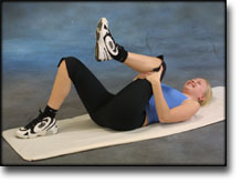
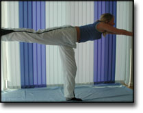
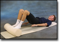
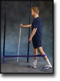
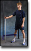
step4
Training ladder for:
FLUID ACCUMULATION IN THE JOINT
(SYNOVITIS/COXITIS)
STEP 4 |
Unlimited: Cycling. Swimming. Running on a smooth surface.
|
||||||||||||||||||||||||||||||||||||||||||||||


|
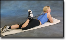
Stretching is carried out in the following way: stretch the muscle group for 3-5 seconds. Relax for 3-5 seconds. The muscle group should subsequently be stretched for 20 seconds. The muscle is allowed to be tender, but must not hurt. Relax for 20 seconds, after which the procedure can be repeated. The time consumed for stretching, coordination and strength training can be altered depending on the training opportunities available and individual requirements. |
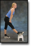
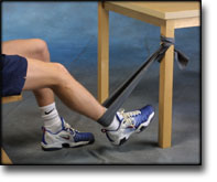
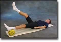
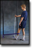
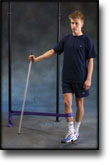
step3
Training ladder for:
FLUID ACCUMULATION IN THE JOINT
(SYNOVITIS/COXITIS)
STEP 3 |
Unlimited: Cycling. Swimming, Light running on a smooth surface.
|
||||||||||||||||||||||||||||||||||||||||||||||
|
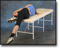
Stretching is carried out in the following way: stretch the muscle group for 3-5 seconds. Relax for 3-5 seconds. The muscle group should subsequently be stretched for 20 seconds. The muscle is allowed to be tender, but must not hurt. Relax for 20 seconds, after which the procedure can be repeated. The time consumed for stretching, coordination and strength training can be altered depending on the training opportunities available and individual requirements. |
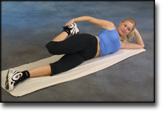
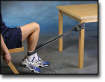
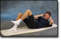
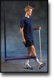
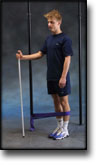
step2
Training ladder for:
FLUID ACCUMULATION IN THE JOINT
(SYNOVITIS/COXITIS)
STEP 2 |
Unlimited: Cycling. Swimming. Jogging on a smooth surface.
|
||||||||||||||||||||||||||||||||||||||||||||||||||||
|
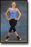
Stretching is carried out in the following way: stretch the muscle group for 3-5 seconds. Relax for 3-5 seconds. The muscle group should subsequently be stretched for 20 seconds. The muscle is allowed to be tender, but must not hurt. Relax for 20 seconds, after which the procedure can be repeated. The time consumed for stretching, coordination and strength training can be altered depending on the training opportunities available and individual requirements. |
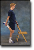
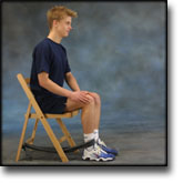
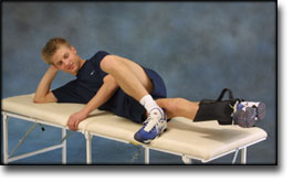
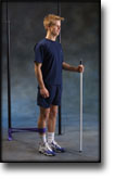
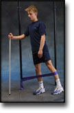
step1
Training ladder for:
FLUID ACCUMULATION IN THE JOINT
(SYNOVITIS/COXITIS)
STEP 1 |
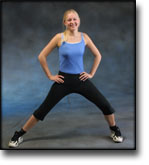
| The indications of time after stretching, coordination training and strength training show the division of time for the respective type of training when training for a period of one hour. The time indications are therefore not a definition of the daily training needs, as the daily training is determined on an individual basis.
|
|||||||||||||||||||||||||||||||||||||||||||||||||||||||
|
Stretching is carried out in the following way: stretch the muscle group for 3-5 seconds. Relax for 3-5 seconds. The muscle group should subsequently be stretched for 20 seconds. The muscle is allowed to be tender, but must not hurt. Relax for 20 seconds, after which the procedure can be repeated. The time consumed for stretching, coordination and strength training can be altered depending on the training opportunities available and individual requirements. |