Kategoriarkiv: Hip

Tendinitis at the ischiatic bone

Untitled Document

Diagnosis: TENDINITIS AT THE ISCHIATIC BONE
(APOPHYSITIS TUBER ISCHIADICUM


Anatomy:
The large posterior thigh muscles (hamstring muscles) have a common muscle tendon fastening on the ischiatic bone (tuber ischiadicum), where a growth zone is also located in children and adolescents. The posterior thigh muscles flex the knee and stretch the hip.

  1. Bursa trochanterica m. glutei maximi
  2. M. gluteus maximus
  3. M. biceps femoris (caput longum)
  4. M. semitendinosus
  5. M. semimembranosus
  6. M. adductor magnus
  7. M. gracilis
  8. M. quadratus femoris
  9. Bursa ischiadica m. glutei maximi

(Photo)

RIGHT GLUTEAL MUSCLES
FROM THE REAR

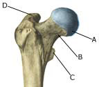
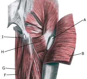
Cause: Inflammation of the tendon fastenings (tendinitis) at the ischiatic bone (tuber ischiadicum) occurs following repeated uniform (over)loads (e.g. running, sprinting) causing microscopic ruptures in the tendon, and especially at the tendon fastening in the growth zone. Inflammation is a warning that the training performed is too strenuous for the muscles in question. In some cases, a single strenuous load can cause a piece of the ischiatic bone to be torn off in the growth zone (article).

Symptoms: Pain in the growth zone in the ischiatic bone can occasionally radiate down into the rear of the thigh. The pain is aggravated when applying pressure on the bone (e.g. sitting position), stretching and activating the posterior thigh muscles (flexing the knee against resistance).

Examination: In slight cases with only minimal tenderness and no discomfort with walking, medical examination is not necessarily required. The extent of the tenderness is, however, not always a mark of the degree of the injury. In cases of more pronounced pain or tenderness, medical examination is required to ensure the correct diagnosis and treatment. The diagnosis is usually made on the basis of a normal medical examination, however, if there is any doubt concerning the diagnosis, this can be confirmed by ultrasound scanning or MR scanning (article). X-ray can be considered if sudden powerful pain is experienced, as this could indicate suspicions of a bone tear.

Treatment: The treatment usually comprises relief, stretching and rehabilitation (article). It can be necessary to re-join the piece of the ischiatic bone which has torn off in the growth zone under surgery in some cases, however, many cases can be successfully treated with relief (article).

Rehabilitation of children and adolescents: INSTRUCTION

Complications: If the treatment does not progress according to plan, it should be considered if the diagnosis is correct or whether complications have arisen. The following should in particular be considered:

Special: Adolescents nearing the end of their teenage years should refer to the adult SportNetDoc under tendonitis at the ischiatic bone.

Fluid accumulation in the hip joint

Untitled Document

Diagnosis: FLUID ACCUMULATION IN THE HIP JOINT
(SYNOVITIS/COXITIS)


Anatomy:
The hip joint consists of the hip socket (acetabulum) and the femoral head (caput femoris). The articular surfaces are coated with a cartilage layer a few mm thick, which reduces the load on the articular surfaces.

Cause: Many repeated loads or one violent load can cause an inflammation of the synovial membrane (synovitis), fluid formation, swelling, restriction of movement and pain in the hip joint. The condition is relatively often seen in children(Drawing).

Symptoms: Smerter i leddet ved bevægelse med belastning. Ofte vil der være bevægelsesindskrænkning ved rotation i hofteleddet.

Examination: It will often be necessary to supplement the ordinary clinical examination with an ultrasound scan, where the fluid in the hip joint can easily be seen (Ultrasonic image).

Treatment: The treatment primarily comprises relief from the pain inducing activity until the swelling has abated. Rehabilitation, within the pain threshold, can subsequently be commenced. In cases of lack of progress with relief the treatment can be supplemented with a medical treatment in the form of rheumatic medicine (NSAID) or by drainage of the joint fluid which should be done with ultrasound guidance.

Rehabilitation of children and adolescents: When the pain has diminished, walking and gradually thereafter running, can be cautiously resumed within the pain threshold, in accordance with rehabilitation of children and adolescents in general.

Complications: lack of progress it should be considered if the diagnosis is correct (article). In particular you should consider a bacterial infection in the joint (pyarthron), which is determined by ultrasound guided draining of the joint fluid, the child hip diseases epifysiolysis capitis femoris (article), Calvé-Legg-Perthes disease (article) and inguinal hernia.

Inguinal hernia

Diagnosis: INGUINAL HERNIA
(HERNIA INGUINALIS)


Anatomy:
The various muscles of the abdominal wall are penetrated by the inguinal canal, which contains nerves (N ilioinguinalis and the genital branch of N genitofemoralis) and in men the spermatic cord (funilicus spermaticus). In women the inguinal canal instead contains a small fibrous ligament. Where the inguinal canal penetrates the abdominal wall weak spots arise (anulus inguinalis superficialis and anulus inguinalis profundus).

  1. M. recti abdominis
  2. Funiculus spermaticus
  3. Ligamentum inguinale
  4. Spina iliaca anterior superior
  5. M. obliquus externus abdominis

Inguinal canal

  1. Anulus inguinalis superficialis
  2. Crus mediale
  3. Funiculus spermaticus et m. cremaster
  4. V. femoralis
  5. Hiatus saphenus
  6. Lig. lacunare
  7. Anulus femoralis
  8. Margo falciformis (cornu superius)
  9. Lig inguinale
  10. Fibrae intercrurales
  11. M. obliquus externus abdominis

Right inguinal canal

Cause: If the abdominal wall becomes too weak, the intestines can be pressed through the weak spots in the abdominal wall causing a hernia.

Symptoms: Pain in the groin, aggravated when coughing. Sometimes a swelling in the groin is visible. Usually the swelling (containing intestine) can be pushed into place. If the swelling is painful and cannot be pressed into place the hernia may be strangulated (which requires acute medical assistance).

Examination: In obvious cases with visible swelling in the groin the diagnosis is easy to make. With lack of swelling in the groin the diagnosis is naturally difficult (article 1). A normal medical examination is usually sufficient in order to make the diagnosis, however, if there is any doubt concerning the diagnosis a MRI-scan or a dynamic ultrasound scan, where weaknesses in the abdominal wall can be detected while increasing the pressure in the abdominal cavity by applying pressure can be performed (article 2), (article 3).

Treatment: With modest discomfort, you can primarily attempt training of the abdominal muscles (article) . If discomfort continues, surgery is advised. If strangulated hernia is suspected you should seek acute medical assistance to evaluate the indication for acute surgery, as there is a risk of lasting damage to the intestine. With uncomplicated surgery, a relief period of approx. two months should be expected before maximal sports activity can be resumed (very dependent on the sport in question).

Rehabilitation of children and adolescents: In normal cases, sports activity will be able to be resumed after a few weeks without special rehabilitation. See: Rehabilitation of children and adolescents in general

Slipping of the femoral head in the growth zone

Diagnosis: SLIPPING OF THE FEMORAL HEADIN THE GROWTH ZONE
(epifysiolyse caput femoris)


Anatomy:
The hip joint consists of the hip socket (acetabulum) and the femoral head (caput femoris). The articular surface is covered with a few mm thick cartilage layer, which reduces the load on the articular surfaces.

  1. Caput femoris
  2. Collum femoris
  3. Trochanter minor
  4. Trochanter major

Femoral head

Cause: For unknown reasons, a slipping of the femoral head in the growth zone can occur in some cases. The condition is mainly seen in boys in the 11-16 years age group (article).

Symptoms: There will only be slight pain initially, with tiredness in the hip joint upon movement and being subjected to load, as well as limping and constriction of movement of the joint. Constriction of movement when rotating the hip joint will often be seen. Increased pain will be felt later on, with more pronounced limping. The pain can occasionally be felt in the knee instead of the hip.

Examination: It is important to undergo a medical examination as soon as possible so that the diagnosis can be made, as this is vital to achieve a good result from the treatment. It will often be necessary to supplement the examination with an x-ray (X-ray) (inclusive of Lauensteins projection), MRI scan or an ultrasound scan (article).

Treatment: Treatment should be commenced as soon as ever possible, and comprises surgery where the articular head is put back into place and fixed if necessary, as well as relief (article).

Rehabilitation of children and adolescents: The type of rehabilitation and load which can be permitted is completely dependant upon the severity of the condition. The rehabilitation should therefore be performed in close cooperation with the doctors controlling the treatment.

Complications: The condition can cause the risk of a lasting injury to the articular head (caput necrose) (article), and degenerative arthritis in the hip joint, as well as shortening of the leg and reduced mobility of the hip joint.

Calve Legg Perthe

Diagnosis: CALVÉ-LEGG-PERTHES DISEASE


Anatomy:
The hip joint consists of the hip socket (acetabulum) and the femoral head (caput femoris). The articular surface is covered with a few mm thick cartilage layer, which reduces the load on the articular surfaces.

  1. Caput femoris
  2. Collum femoris
  3. Trochanter minor
  4. Trochanter major

Femoral head

Cause: For unknown reasons, a slow collapse of the femoral head (aseptic necrosis of the bone) can occur, resulting in a disintegration and flattening of the femoral head and consequently irritation in the hip joint. However, secondhand smoke is recognised as playing a role  (article). The condition is mainly seen in children in the 3-11 age group, with boys being affected three times more often than girls. 10% of cases are on both sides of the hip.

Symptoms: There will only be slight pain initially, with tiredness in the hip joint upon movement and being subjected to load, as well as limping and constriction of movement of the joint. Constriction of movement when rotating the hip joint will often be observed. Increased pain will be felt later on, with more pronounced limping due to the shortening of the leg. The pain can occasionally be felt in the knee instead of the hip.

Examination: It is important to undergo a medical examination as soon as possible so that the diagnosis can be made, as this is vital to achieve a good result from the treatment. It will often be necessary to supplement the examination with an ultrasound scan (Ultrasonic image), where bone change and fluid in the hip joint can clearly be identified (article) and possibly also x-ray examination where the late bone change can be seen (X-ray).

Treatment: Treatment should be commenced as soon as ever possible, and primarily comprises intensive relief, with possible use of a wheel chair. The period of relief can last 1-2 years. An operative correction can be necessary if the disease leaves considerable damage (article).

Rehabilitation of children and adolescents: The type of rehabilitation and load which can be permitted is completed dependant upon the severity of the disease. The rehabilitation should therefore be performed in close cooperation with the doctors controlling the treatment

Complications: The condition can cause the risk of shortening of the leg and degenerative arthritis in the hip joint. There are, however, good chances for the articular head to heal to its normal shape, especially for the youngest children.