Kategoriarkiv: Shinbone

Inflammation of the outer shin bone


Diagnosis: INFLAMMATION OF THE OUTER SHIN BONE
(LATERAL TIBIAL PERIOSTITIS)


Anatomy:
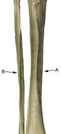
The calf muscles are divided in three muscle groups (compartments) by powerful muscle membranes (fascies). A forward, an exterior and a rear muscle group (which is divided in a superficial and a deep part). The large rear muscle groups’ muscle membrane is fastened on the inner edge of the shin bone, while the forward muscle group is fastened on the external edge of the shin bone (photo).

Cause: In cases of repeated uniform loads, a stress related inflammation, can occur where the muscle membrane fastens on the edge of the shin bone. The condition is most often seen in athletes who jump a lot and with frequent change of running surface or shoes.

Symptoms: Pain upon applying pressure, particularly on the exterior edge of the shin bone and upon straining, and is aggravated when the foot is bent upwards against resistance (extension). Occasionally an irregular bone edge can be felt on the shin bone.

Acute treatment:

Examination: In light cases medical examination is not necessarily required. In cases with more pronounced pain or lack of progress despite relief, a medical examination is recommended to ensure the diagnosis and rule out amongst other things a stress fracture. A medical examination is usually sufficient in order to make the diagnosis. It can be necessary to supplement with X-ray, Scintigraphy, ultrasonography or MRI (article).

Treatment: The treatment involves relief, stretching and slow rehabilitation. It is imperative that there are good shock absorbing soles in the shoes.

Rehabilitation of children and adolescents: INSTRUCTION

Complications: If the course does not progress smoothly, you should be medically re-examined to ensure that the diagnosis is correct, and that complications have not arisen in the form of stress fracture.

Special: Shock absorbing shoes or inlays will reduce the load.


Inflammation of the inner shin bone

Diagnosis: INFLAMMATION OF THE INNER SHIN BONE
(MEDIAL TIBIAL PERIOSTITIS, “SHIN SPLINT”)


Anatomy:
The calf muscles are divided in three muscle groups (compartments) by powerful muscle membranes (fascies). A forward, an exterior and a rear muscle group (which is divided into a superficial and a deep part). The large rear muscle groups’ muscle membrane is fastened on the inner edge of the shin bone (tibia), while the forward muscle group is fastened on the external edge of the shin bone (photo).

Cause: In cases of repeated uniform loads, a stress related inflammation, can occur where the muscle membrane fastens on the edge of the shin bone. The condition is most often seen in athletes with a tendency to rotate the foot outwards (hyperpronation) or with a high foot arch. Frequent change of running surface or shoes increases the risk.

Symptoms: Pain on the inner edge of the shin bone, aggravated upon applying pressure, load (running) and stretching in the foot joint against resistance (flexion). The pain is often localized to the lower part of the shin bone. Sometimes an irregular bone edge can be felt on the shin bone.

Acute treatment:

Examination: In light cases medical examination is not necessarily required. In cases with more pronounced pain or lack of progress despite relief, medical examination is recommended to ensure the diagnosis and rule out amongst other things a stress fracture. A medical examination is usually sufficient in order to make the diagnosis. In some cases it may be necessary to supplement with X-rays, scintigraphy or ultrasound scanning.

Treatment: The treatment comprises relief, stretching and slow rehabilitation. It is imperative that there are good shock absorbing soles in the shoes.

Rehabilitation of children and adolescents: INSTRUCTION

Complications: If the course does not progress smoothly, you should be medically re-examined to ensure that the diagnosis is correct, and that complication have not arisen in the form of stress fracture.

Special: Shock absorbing shoes or inlays will reduce the load.

Bone fracture

Diagnosis: BONE FRACTURE OF THE SHIN BONE
(FRACTURE)


Anatomy:
The bones in the lower leg comprise the shin bone (tibia) and the calf bone (fibula).

  1. Tibia
  2. Fibula

SHIN BONE FROM THE FRONT

Cause: Violent loads in the form of a blow or twist can cause a fracture on the shin-bone as well as the calf bone (article). Fracture of the fibula is often seen after a kick on the outer side of the shin bone.

Symptoms: Pain upon applying pressure (direct and indirect tenderness) and when under load (walking and running).

Acute treatment: Click here.

Examination: X-ray.

Treatment: The treatment comprises relief and bandaging. In some cases surgery is necessary depending on the type of fracture and any displacement of the fracture-surfaces (article).

Rehabilitation of children and adolescents: Which loads and rehabilitation that can be permitted is completely dependent upon the severity and the treatment of the fracture. It is therefore important that the rehabilitation is performed in close cooperation with the doctors controlling the treatment. Approximately six months’ rehabilitation must be expected before shin bone fractures allow resumption of maximum load, and approximately 3 months’ rehabilitation before calf bone fractures allow maximum load.

Plastic bandage: Individual plastic bandages can be manufactured for use during sports activity after bone fractures. Individual plastic bandages are particularly well-suited after a fracture of the fibula.

Complications: If progress is not smooth you should be medically re-examined to ensure that the fracture is healing according to plan. In some cases a false joint can be formed (pseudoarthrosis) (article), requiring surgical treatment.