Kategoriarkiv: Foot, Achilles

KONDITION

step1

Training ladder for:
INFLAMMATION OF THE ACHILLES TENDON ATTACHMENT
(Achilles enthesopathy)

STEP 1

The indications of time after stretching, coordination training and strength training show the division of time for the respective type of training when training for a period of one hour. The time indications are therefore not a definition of the daily training needs, as the daily training is determined on an individual basis.

KONDITION
Unlimited: Cycling. Swimming.

UDSPÆNDING
(15 min)

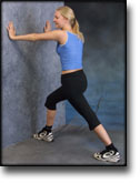
Stand with the injured leg stretched backwards with the toes facing front. Slowly bend the knee so that the calf muscles become increasingly stretched. Hold the position for 20 seconds and relax for 20 seconds before repeating.

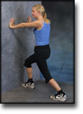
Stand with the injured leg outstretched with the ankle joint bent up against the wall. Press your abdomen against the wall so that the calf muscles become increasingly stretched. Hold the position for 20 seconds and relax for 20 seconds before repeating.

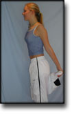
Stand on the good leg while gaining support on the wall with the hand on the same side. Bend the knee of the injured leg and draw the heel towards the buttocks. Take hold of the big toe side of the foot and draw slowly upwards so that the ankle joint is stretched to the maximum and drawn slightly outwards so that increased stretching is experienced on the inside of the shin bone. Hold the position for 20 seconds and relax for 20 seconds before repeating.

Stand on the good leg while gaining support from the wall with the opposite hand. Bend the knee of the injured leg and draw the heel towards the buttocks. Take hold of the little toe side of the foot and draw slowly upwards so that the ankle joint is stretched to the maximum and drawn over the good leg so that increased stretching is experienced on the outer side of the shin bone. Hold the position for 20 seconds and relax for 20 seconds before repeating.

KOORDINATION
(5 min)

Seesaw. Balance on two legs, possibly using a hand as support against the wall, balancing subsequently on one leg without support. Look straight ahead and keep knees bent.

STYRKE
(40 min)

Sit on the floor. Tip the foot up and down with elastic under the forefoot so that the elastic becomes taut.

Sit on the floor. Tip the foot up and down with elastic on top of the forefoot so that the elastic becomes taut.

Sit on the floor. Tip the foot from side to side with elastic on the outer side of the foot, without moving the knee.

Sit on the floor. Tip the foot from side to side with elastic on the inner side of the foot, without moving the knee.

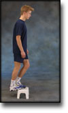
Stand with both forefeet on a stool or doorstep with the heel out over the edge. Slowly rise up on to your toes with your weight on the healthy leg. Go slowly down on the injured leg as far as you can go. Use the healthy leg to rise up on to your toes again. The exercise should be performed with stretched, as well as bent knee. Wearing a rucksack and gradually increasing the ballast in the rucksack can increase the load.

Stretching is carried out in the following way: stretch the muscle group for 3-5 seconds. Relax for 3-5 seconds. The muscle group should subsequently be stretched for 20 seconds. The muscle is allowed to be tender, but must not hurt. Relax for 20 seconds, after which the procedure can be repeated. The time consumed for stretching, coordination and strength training can be altered depending on the training opportunities available and individual requirements.

treatment-article1

SportNetDoc

Chronic Achilles tendon pain treated with eccentric calf-muscle training.

Fahlstrom M, Jonsson P, Lorentzon R, Alfredson H. Knee Surg Sports Traumatol Arthrosc. 2003 Sep;11(5):327-33.

Injuries involving the Achilles tendon and manifested as chronic tendon pain are common, especially among recreational athletes. In a pilot study on a small group of patients with chronic painful mid-portion Achilles tendinosis, eccentric calf-muscle training was shown to give good clinical results. The aim of this prospective study was to investigate if the previously achieved good clinical results could be reproduced in a larger group of patients, and also to investigate the effects of eccentric calf-muscle training in patients with chronic insertional Achilles tendon pain. Seventy-eight consecutive patients, having chronic painful Achilles tendinosis at the mid-portion (2-6 cm level) in a total of 101 tendons (55 unilateral and 23 bilateral), and thirty consecutive patients with chronic insertional Achilles tendon pain in 31 tendons (29 unilateral and one bilateral) were treated with eccentric calf-muscle training for 12 weeks. Most patients were recreational athletes. Evaluation of the amount of tendon pain during activity was recorded on a visual analogue scale (VAS), before and after treatment. In 90 of the 101 Achilles tendons (89%) with chronic painful mid-portion Achilles tendinosis, treatment was satisfactory and the patients were back on their pre-injury activity level after the 12-week training regimen. In these patients, the amount of pain during activity, registered on the VAS-scale (mean+/-SD), decreased significantly from 66.8+/-19.4 to 10.2+/-13.7. On the contrary, in only ten of the tendons (32%) with chronic insertional Achilles tendon pain was treatment satisfactory, with a significant decrease on the VAS-scale (mean+/-SD), from 68.3+/-7.0 to 13.3+/-13.2. Our conclusion is that treatment with eccentric calf-muscle training produced good clinical results in patients with chronic painful mid-portion Achilles tendinosis, but not in patients with chronic insertional Achilles tendon pain.

treatment-article3

SportNetDoc

Local corticosteroid injection in sport: review of literature and guidelines for treatment.

Fredberg U. Scand J Med Sci Sports 1997 Jun;7(3):131-9.

The risks and benefits of local injection therapy of overuse sports injuries with corticosteroids are reviewed here. Injection of corticosteroid inside the tendon has a deleterious effect on the tendon tissue and should be unanimously condemned. No reliable proof exists of the deleterious effects of peritendinous injections. Too many conclusions in the literature are based on poor scientific evidence and it is just the reiteration of a dogma if all steroid injections are abandoned. The corticosteroids represent an adjuvant treatment in the overall management of sports injuries: basic treatment is ‘active’ rest and graduated rehabilitation within the limits of pain. With proper indications there are only few and trivial complications that may occur with corticosteroid injections. Guidelines for proper local injection therapy with corticosteroids are given.

treatment-article2

SportNetDoc

Local corticosteroid injection in sport: review of literature and guidelines for treatment.

Fredberg U. Scand J Med Sci Sports 1997 Jun;7(3):131-9.

The risks and benefits of local injection therapy of overuse sports injuries with corticosteroids are reviewed here. Injection of corticosteroid inside the tendon has a deleterious effect on the tendon tissue and should be unanimously condemned. No reliable proof exists of the deleterious effects of peritendinous injections. Too many conclusions in the literature are based on poor scientific evidence and it is just the reiteration of a dogma if all steroid injections are abandoned. The corticosteroids represent an adjuvant treatment in the overall management of sports injuries: basic treatment is ‘active’ rest and graduated rehabilitation within the limits of pain. With proper indications there are only few and trivial complications that may occur with corticosteroid injections. Guidelines for proper local injection therapy with corticosteroids are given.

special-article

SportNetDoc

Significance of ultrasonographically detected asymptomatic tendinosis in the patellar and achilles tendons of elite soccer players: a longitudinal study.

Fredberg U, Bolvig L. Am J Sports Med 2002 Jul-Aug;30(4):488-91

BACKGROUND: Chronic tendinosis of the Achilles or patellar tendons, or both, is one of the most frequent and severe conditions that affects athletes in sports such as soccer. It can often end an athlete’s sports activity. HYPOTHESIS: Ultrasonography of asymptomatic tendons can be used to predict which athletes will develop tendon symptoms. STUDY DESIGN: Longitudinal study. METHODS: Using ultrasonography of the ankle and knee, we examined 54 elite soccer players in the top Danish soccer league before and after a single season (in January and December). RESULTS: At the start of the season, ultrasonography had revealed abnormalities in 29% of those examined. Eighteen percent (18 of 98 tendons) were observed to have abnormal sonographic findings in the patellar tendon at the initial examination in January. These athletes were found to have a 17% risk of developing symptomatic jumper’s knee during the 12-month season. Eleven percent (11 of 96 tendons) were observed to have abnormal sonographic findings in the Achilles tendon at the initial examination; it was calculated that they had a 45% risk of developing symptoms of Achilles tendinosis. Only one of the players with normal tendons in January actually developed symptoms by the end of the season. CONCLUSIONS: For the first time it is now possible to identify risk factors for the development of serious tendon disorders in asymptomatic athletes. Future studies may be directed at developing preventive treatment to reduce the risk of chronic, therapy-resistant symptoms of tendinosis and ruptures.

treatment-article2

SportNetDoc

Surgical management of Achilles tendon overuse injuries. A long-term follow-up study.

Schepsis AA, Wagner C, Leach RE. Am J Sports Med 1994 Sep-Oct;22(5):611-9.

We studied 79 cases of surgically treated Achilles tendon overuse injuries in 66 patients. Fifty-three (80%) of these patients were competitive or serious recreational runners operated on between 1978 and 1991. There were 49 men and 17 women with a mean age of 33 years (range, 17 to 59). The cases were divided into surgical subgroups based on their site of primary symptoms and abnormalities: paratenonitis (23), tendinosis (partial rupture or degeneration) (15), retrocalcaneal bursitis (24), insertional tendinitis (7), and combined abnormalities (10). Followup included a comprehensive patient questionnaire and office examination. There were 79% satisfactory (51% excellent, 28% good) and 21% unsatisfactory (17% fair, 4% poor) results. The percentages of satisfactory results in the paratenonitis group (87%) were best and those in the tendinosis group were the worst (67%). Satisfactory results were obtained in 75% of the patients with retrocalcaneal bursitis and 86% with insertional tendinitis. Seven of the 45 cases with longer than 5-year followup with initially satisfactory results deteriorated with time and required reoperation (16%). Of these, 4 were in the tendinosis group, 2 had retrocalcaneal bursitis, and 1 had paratenonitis. One of the 34 patients followed less than 5 years required reoperation.

treatment-article1

SportNetDoc

Chronic Achilles tendinosis: recommendations for treatment and prevention.

Alfredson H, Lorentzon R. Sports Med 2000 Feb;29(2):135-46.

Chronic Achilles tendinosis is a condition with an unknown aetiology and pathogenesis that is often, but not always, associated with pain during loading of the Achilles tendon. Histologically, there are no inflammatory cells, but increased amounts of interfibrillar glycosaminoglycans and changes in the collagen fibre structure and arrangement are seen. In situ microdialysis has confirmed the absence of inflammation. It is a condition that is most often seen among recreational male runners aged between 35 and 45 years, and it is most often considered to be associated with overuse. However, this condition is also seen in patients with a sedentary lifestyle. Chronic Achilles tendinosis is considered a troublesome injury to treat. Nonsurgical treatment most often includes a combination of rest, NSAIDs, correction of malalignments, and stretching and strengthening exercises, but there is sparse scientific evidence supporting the use of most proposed treatment regimens. It has been stated that, in general, nonsurgical treatment is not successful and surgical treatment is required in about 25% of patients. However, in a recent prospective study, treatment with heavy load eccentric calf muscle training showed very promising results and may possibly reduce the need for surgical treatment of tendinosis located in the midportion of the Achilles tendon. The short term results after surgical treatment are frequently very good, but in the few studies with long term follow-up there are signs of a possible deterioration with time. Calf muscle strength takes a long time to recover and, furthermore, a prolonged progressive calcaneal bone loss has been shown on the operated side up to 1 year after surgical treatment.

treatment-article4

SportNetDoc

Ultrasonography as a tool for diagnosis, guidance of local steroid injection and, together with pressure algometry, monitoring of the treatment of athletes with chronic jumper’s knee and Achilles tendinitis: a randomized, double-blind, placebo-controlled study.

Fredberg U, Bolvig L, Pfeiffer-Jensen M, Clemmensen D, Jakobsen BW, Stengaard-Pedersen K. Scand J Rheumatol. 2004;33(2):94-101.

BACKGROUND: The diagnosis of Achilles and patella tendinitis has until recently been based on clinical examination, and treatment with local steroid injection has been given blindly. This is the first randomized, double blind, placebo-controlled study of local steroid injection in athletes with chronic tendinitis, which used ultrasonography to increase diagnostic accuracy, to guide the correct placement of local steroid and, conjunctively with pressure algometry, to objectify and monitor the results of treatment. METHOD: Forty-eight athletes each with severe symptomatic tendinitis of a patellar (24) or Achilles tendon (24) for more than 6 months, whose conditions were confirmed ultrasonographically, and who all failed conservative treatment (rehabilitation) were included in this double-blind, placebo-controlled study and treated with three ultrasonographically guided peritendinous injections of steroid or placebo. RESULTS: The conditions of only one-third of the referred athletes with clinically suspected tendinitis were confirmed by ultrasonographic examination. The ultrasonographically guided peritendinous injection of steroid had a significant effect in reducing pain and thickening of tendons. CONCLUSION: Ultrasonography should be used in the future to assure precise diagnosis and to guide the peritendinous injection of steroid in chronic Achilles and patella tendinitis. Ultrasonography and pressure algometry are recommended as objective methods for monitoring the effect of treatment. Ultrasonographically guided injection of long-acting steroid can normalize the ultrasonographic pathological lesions in the Achilles and patellar tendons, and has a dramatic clinical effect but when combined with aggressive rehabilitation with running after a few days, many will have relapse of symptoms within 6 months (see the article – 1,5 mb).

KONDITION

step4

Training ladder for:
INFLAMMATION OF THE ACHILLES TENDON
(TENDINITIS ACHILII)

STEP 4

KONDITION
Unlimited: Cycling. Swimming. Running with increasing distance on a soft surface.

UDSPÆNDING
(15 min)

Stand with the injured leg stretched backwards with the toes facing front. Slowly bend the knee so that the calf muscles become increasingly stretched. Hold the position for 20 seconds and relax for 20 seconds before repeating.

Stand with the injured leg outstretched with the ankle joint bent up against the wall. Press your abdomen against the wall so that the calf muscles become increasingly stretched. Hold the position for 20 seconds and relax for 20 seconds before repeating.

Stand on the good leg while gaining support on the wall with the hand on the same side. Bend the knee of the injured leg and draw the heel towards the buttocks. Take hold of the big toe side of the foot and draw slowly upwards so that the ankle joint is stretched to the maximum and drawn slightly outwards so that increased stretching is experienced on the inside of the shin bone. Hold the position for 20 seconds and relax for 20 seconds before repeating.

Stand on the good leg while gaining support from the wall with the opposite hand. Bend the knee of the injured leg and draw the heel towards the buttocks. Take hold of the little toe side of the foot and draw slowly upwards so that the ankle joint is stretched to the maximum and drawn over the good leg so that increased stretching is experienced on the outer side of the shin bone. Hold the position for 20 seconds and relax for 20 seconds before repeating.

KOORDINATION
(5 min)

Stand on the leg to be trained. Take-off and land on the same leg.

Seesaw. Balance on two legs, possibly using a hand as support against the wall, balancing subsequently on one leg without support. Look straight ahead and keep knees bent.

STYRKE
(40 min)

Sit on the floor. Tip the foot up and down with elastic under the forefoot so that the elastic becomes taut.

Sit on the floor. Tip the foot up and down with elastic on top of the forefoot so that the elastic becomes taut.

Sit on the floor. Tip the foot from side to side with elastic on the outer side of the foot, without moving the knee.

Sit on the floor. Tip the foot from side to side with elastic on the inner side of the foot, without moving the knee.

Stand on a soft surface. Rise slowly up on tiptoe and go down again.

Stand behind a chair. Rise slowly up on tiptoe and go down again.

Stand with both forefeet on a stool or doorstep with the heel out over the edge. Slowly rise up on to your toes with your weight on the healthy leg. Go slowly down on the injured leg as far as you can go. Use the healthy leg to rise up on to your toes again. The exercise should be performed with stretched, as well as bent knee. Wearing a rucksack and gradually increasing the ballast in the rucksack can increase the load.

Stand with feet together. Using the ankle joint to take off, hop approx. 5 cm and land on both feet. The exercise should be done on one leg when you are able to do it without discomfort using both legs.

Stretching is carried out in the following way: stretch the muscle group for 3-5 seconds. Relax for 3-5 seconds. The muscle group should subsequently be stretched for 20 seconds. The muscle is allowed to be tender, but must not hurt. Relax for 20 seconds, after which the procedure can be repeated. The time consumed for stretching, coordination and strength training can be altered depending on the training opportunities available and individual requirements.

KONDITION

step3

Training ladder for:
INFLAMMATION OF THE ACHILLES TENDON
(TENDINITIS ACHILII)

STEP 3

KONDITION
Unlimited: Cycling. Swimming. Light jogging on a soft surface.

UDSPÆNDING
(15 min)

Stand with the injured leg stretched backwards with the toes facing front. Slowly bend the knee so that the calf muscles become increasingly stretched. Hold the position for 20 seconds and relax for 20 seconds before repeating.

Stand with the injured leg outstretched with the ankle joint bent up against the wall. Press your abdomen against the wall so that the calf muscles become increasingly stretched. Hold the position for 20 seconds and relax for 20 seconds before repeating.

Stand on the good leg while gaining support on the wall with the hand on the same side. Bend the knee of the injured leg and draw the heel towards the buttocks. Take hold of the big toe side of the foot and draw slowly upwards so that the ankle joint is stretched to the maximum and drawn slightly outwards so that increased stretching is experienced on the inside of the shin bone. Hold the position for 20 seconds and relax for 20 seconds before repeating.

Stand on the good leg while gaining support from the wall with the opposite hand. Bend the knee of the injured leg and draw the heel towards the buttocks. Take hold of the little toe side of the foot and draw slowly upwards so that the ankle joint is stretched to the maximum and drawn over the good leg so that increased stretching is experienced on the outer side of the shin bone. Hold the position for 20 seconds and relax for 20 seconds before repeating.

KOORDINATION
(5 min)

Stand on one leg. Play the ball up against the wall.

Seesaw. Balance on two legs, possibly using a hand as support against the wall, balancing subsequently on one leg without support. Look straight ahead and keep knees bent.

STYRKE
(40 min)

Sit on the floor. Tip the foot up and down with elastic under the forefoot so that the elastic becomes taut.

Sit on the floor. Tip the foot up and down with elastic on top of the forefoot so that the elastic becomes taut.

Sit on the floor. Tip the foot from side to side with elastic on the outer side of the foot, without moving the knee.

Sit on the floor. Tip the foot from side to side with elastic on the inner side of the foot, without moving the knee.

Stand on a soft surface. Rise slowly up on tiptoe and go down again.

Stand behind a chair. Rise slowly up on tiptoe and go down again.

Stand with both forefeet on a stool or doorstep with the heel out over the edge. Slowly rise up on to your toes with your weight on the healthy leg. Go slowly down on the injured leg as far as you can go. Use the healthy leg to rise up on to your toes again. The exercise should be performed with stretched, as well as bent knee. Wearing a rucksack and gradually increasing the ballast in the rucksack can increase the load.

Stretching is carried out in the following way: stretch the muscle group for 3-5 seconds. Relax for 3-5 seconds. The muscle group should subsequently be stretched for 20 seconds. The muscle is allowed to be tender, but must not hurt. Relax for 20 seconds, after which the procedure can be repeated. The time consumed for stretching, coordination and strength training can be altered depending on the training opportunities available and individual requirements.