Kategoriarkiv: foot, front

tape-instruction

Tapening


Type: TOE NAIL:

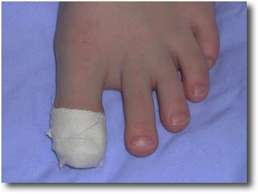
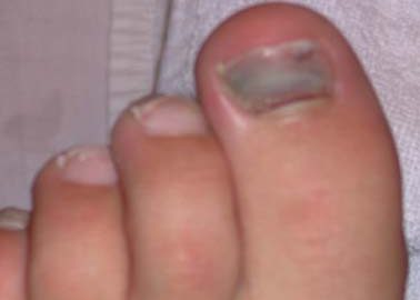
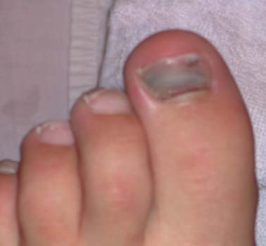
Objective: To secure a partially loose big toe nail following bleeding under the nail.

Application: One or two strips of tape are applied around the toe and toe nail. The tape should be fixed so tightly as to ensure that the nail is pressed against the toe, without causing the toe to sleep.

treatment-article

SportNetDoc

Subungual haematomas: is simple trephining enough?

Meek S, White M. J Accid Emerg Med 1998 Jul;15(4):269-71.

OBJECTIVES: To determine the outcome of treating subungual haematomas by simple trephining, in terms of nail appearance and function, and to identify factors associated with a poor outcome. METHODS: Patients with subungual haematomas attending an accident and emergency department over a 12 month period were identified; 5-13 months after injury a telephone and postal survey of patient’s assessment of their own outcome was made using a proforma. Outcome was graded according to published criteria. RESULTS: 123 patients with 127 subungual haematomas were identified and 74% were followed up; 85% achieved an “excellent” or “very good” result, with 67% reporting no residual abnormalities. A “poor” outcome occurred in only 2% owing to nail splitting. No correlation was found between adverse outcome and haematoma size, presence of fracture, or infection. CONCLUSIONS: Treatment of subungual haematomas by simple trephining gives an acceptable result in the majority of patients and those with a poor outcome cannot be predicted at presentation. The findings suggest that only simple trephining for symptomatic relief is required for subungual haematomas and that aggressive surgical treatment is unnecessary.

cause-article

SportNetDoc

Trauma to the nail unit including occupational sports injuries.

Mortimer PS, Dawber RP. Dermatol Clin 1985 Jul;3(3):415-20.

Trauma to the nail unit is one of the most common causes of onychodystrophy and may be acute (a single overwhelming injury) or chronic (repeated minor injury). Immediate effects of acute trauma include splinter hemorrhages, subungual hematomas, and nail shedding. Delayed deformities include splits and ridges, pterygium, hook nails, pigment bands, and ectopic nail. Also discussed are the results of chronic trauma, including self-inflicted injury and repeated indirect injury.

complication-article

SportNetDoc

Hallucal sesamoid pain: causes and surgical treatment.

Richardson EG. J Am Acad Orthop Surg 1999 Jul-Aug;7(4):270-8.

The hallucal sesamoids, although small and seemingly insignificant, play an important role in the function of the great toe by absorbing weight-bearing pressure, reducing friction, and protecting tendons. However, the functional complexity and anatomic location of these small bones make them vulnerable to injury from shear and loading forces. Injury to the hallucal sesamoids can cause incapacitating pain, which can be devastating to an athlete. Although traumatic injuries usually can be diagnosed easily, other pathologic conditions may be overlooked. Careful physical and radiologic examinations are necessary to determine the cause of pain and allow a recommendation of the optimal treatment. Surgical treatment may include partial or complete resection of the sesamoid, shaving of a prominent tibial sesamoid, or autogenous bone grafting for nonunion. Excision of both sesamoids should be avoided if possible.

examination-article

SportNetDoc

Disorders of the hallux sesamoid complex: MR features.

Karasick D, Schweitzer ME. Skeletal Radiol 1998 Aug;27(8):411-8.

Numerous painful conditions can affect the first metatarsophalangeal-sesamoid joint complex. Symptoms can be of sudden or insidious onset, and be of acute or chronic duration. Although conventional radiography is recognized as the initial diagnostic procedure for these symptoms, there is often a need to proceed to MR imaging. MR imaging is sensitive and can be utilized in the investigation of the hallux sesamoid complex to differentiate soft tissue from osseous pathology. Synovitis, tendonitis, and bursitis can be distinguished from bony abnormalities such as sesamoid fracture, avascular necrosis, and osteomyelitis. An understanding of MR imaging features and techniques will result in the highest diagnostic yield. Early and accurate diagnosis of sesamoid complex disorders can guide the physician to the appropriate clinical management and prevent potentially harmful longstanding joint dysfunction.

treatment-article2

SportNetDoc

The treatment of rigid hallux using Swanson’s silastic implant (single and double stem). Clinical, radiological and podobarographic review with a 16-year maximum follow-up.

Mahieu C, Chaput A, Bouillet R. Acta Orthop Belg 1992;58(3):314-24.

Twenty-eight cases of hallux rigidus treated with the Swanson Silastic implant (single and double stem) were reviewed, with an 8-year follow-up. The skin complication rate was significant. Long-term patient satisfaction was good. Radiologic findings were alarming: implants seem to wear out quickly on the articular side, and granulomatous reactions develop around the stems. Dynamic pedobarography shows decreased pressure under the first ray, with transfer of the weight to the midmetatarsal heads, sometimes on the external edge of the foot. The authors review the international literature on the etiology, pathology and treatment of hallux rigidus. Surgical indications and techniques are specified.

treatment-article1

SportNetDoc

Outcomes in hallux rigidus patients treated nonoperatively: a long-term follow-up study.

Smith RW, Katchis SD, Ayson LC. Foot Ankle Int 2000 Nov;21(11):906-13.

The purpose of this study was to analyze radiographic outcome and patient satisfaction in non-operative care of hallux rigidus. Twenty-two patients representing 24 feet were surveyed and radiographed. Average follow-up was 14.4 years (range, 12-19 years). In 75% (18/24) of the feet, the patients would “still chose not to have surgery” if they had to make the decision again. The pain remained about the same in 22 feet, improved with time in one, and became worse in one. The most common reason given for not having surgery was that the pain was not severe enough. The most common type of self-care was a shoe with an “ample toe box.” More patients benefited from a stiff sole than a soft sole, but the majority of patients did not cite the sole of the shoe as being important. There was measurable loss of cartilage space radiographically over time in 16 of 24 feet, and in eight of the 16 feet, the loss of cartilage space was dramatic. The majority of hallux rigidus patients rated their pain as staying the same over a twelve-year period, despite significant deterioration of joint space noted radiographically.

examination-article1

SportNetDoc

Hallux valgus and hallux rigidus: MRI findings.

Schweitzer ME, Maheshwari S, Shabshin N. Clin Imaging 1999 Nov-Dec;23(6):397-402.

The purpose of this article is to describe the MR findings of Hallux Valgus (HV) and Hallux Rigidus (HR). Twenty-four patients (11 with HV, 4 with HR, and 9 with both HV and HR) were studied at 1.5 Tesla MRI. Two separate observers evaluated the first ray blindly for the following signs: sesamoid position, sesamoid proliferation, hypertrophy of the median eminence, presence of a lateral facet, presence of an adventitial bursa, shape of the first metatarsal head, relative length of the first metatarsal, joint space loss, osteophytes (dorsalor lateral), marrow edema, geodes, subchondral sclerosis, intra-articular ossicle, and pes planus. The most common findings observed in HV were a hypertrophic medial eminence (95%), sesamoid proliferation (90%) and adventitial bursitis (70%). The most common findings observed in HR were osteophytes (77% and 69%), geodes, and marrow edema. We conclude that traditional routine radiograph signs of HV and HR may be applied to MR images.