Kategoriarkiv: Inflammation of the hollow tendon

KONDITION

step4

Training ladder for:
INFLAMMATION OF THE HOLLOW FOOT TENDON
(FASCIITIS PLANTARIS)

STEP 4

KONDITION
Unlimited: Cycling. Swimming.

UDSPÆNDING
(10 min)

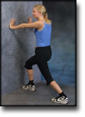
Stand with the injured leg stretched backwards with the toes facing front. Slowly bend the knee so that the calf muscles become increasingly stretched. Hold the position for 20 seconds and relax for 20 seconds before repeating.

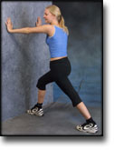
Stand with the injured leg outstretched with the ankle joint bent up against the wall. Press your abdomen against the wall so that the calf muscles become increasingly stretched. Hold the position for 20 seconds and relax for 20 seconds before repeating.

KOORDINATION
(5 min)

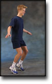
Seesaw. Balance on two legs, possibly using a hand as support against the wall, balancing subsequently on one leg without support. Look straight ahead and keep knees bent.

STYRKE
(45 min)

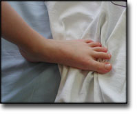
Sit with your bare toes on a tea towel and curl the tea towel together using your toes.

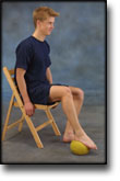
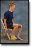
Sit with a tennis ball under the foot. Roll the ball backwards and forwards and from side to side.

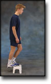
Stand with both forefeet on a stool or doorstep with the heel out over the edge. Slowly rise up on to your toes with your weight on the healthy leg. Go slowly down on the injured leg as far as you can go. Use the healthy leg to rise up on to your toes again. The exercise should be performed with stretched, as well as bent knee. Wearing a rucksack and gradually increasing the ballast in the rucksack can increase the load.

Stand on a soft surface. Rise slowly up on tiptoe and go down again.

Stand on your toes with bent knees. Place your weight forward on the toes and keep your balance.

Stretching is carried out in the following way: stretch the muscle group for 3-5 seconds. Relax for 3-5 seconds. The muscle group should subsequently be stretched for 20 seconds. The muscle is allowed to be tender, but must not hurt. Relax for 20 seconds, after which the procedure can be repeated. The time consumed for stretching, coordination and strength training can be altered depending on the training opportunities available and individual requirements.

KONDITION

step3

Training ladder for:
INFLAMMATION OF THE HOLLOW FOOT TENDON
(FASCIITIS PLANTARIS)

STEP 3

KONDITION
Unlimited: Cycling. Swimming.

UDSPÆNDING
(10 min)

Stand with the injured leg stretched backwards with the toes facing front. Slowly bend the knee so that the calf muscles become increasingly stretched. Hold the position for 20 seconds and relax for 20 seconds before repeating.

Stand with the injured leg outstretched with the ankle joint bent up against the wall. Press your abdomen against the wall so that the calf muscles become increasingly stretched. Hold the position for 20 seconds and relax for 20 seconds before repeating.

KOORDINATION
(5 min)

Seesaw. Balance on two legs, possibly using a hand as support against the wall, balancing subsequently on one leg without support. Look straight ahead and keep knees bent.

STYRKE
(45 min)

Sit with your bare toes on a tea towel and curl the tea towel together using your toes.

Sit with a tennis ball under the foot. Roll the ball backwards and forwards and from side to side.

Stand with both forefeet on a stool or doorstep with the heel out over the edge. Slowly rise up on to your toes with your weight on the healthy leg. Go slowly down on the injured leg as far as you can go. Use the healthy leg to rise up on to your toes again. The exercise should be performed with stretched, as well as bent knee. Wearing a rucksack and gradually increasing the ballast in the rucksack can increase the load.

Stand on a soft surface. Rise slowly up on tiptoe and go down again.

Stand on your toes with bent knees. Place your weight forward on the toes and keep your balance.

Stretching is carried out in the following way: stretch the muscle group for 3-5 seconds. Relax for 3-5 seconds. The muscle group should subsequently be stretched for 20 seconds. The muscle is allowed to be tender, but must not hurt. Relax for 20 seconds, after which the procedure can be repeated. The time consumed for stretching, coordination and strength training can be altered depending on the training opportunities available and individual requirements.

KONDITION

step2

Training ladder for:
INFLAMMATION OF THE HOLLOW FOOT TENDON
(FASCIITIS PLANTARIS)

STEP 2

KONDITION
Unlimited: Cycling. Swimming.

UDSPÆNDING
(10 min)

Stand with the injured leg stretched backwards with the toes facing front. Slowly bend the knee so that the calf muscles become increasingly stretched. Hold the position for 20 seconds and relax for 20 seconds before repeating.

Stand with the injured leg outstretched with the ankle joint bent up against the wall. Press your abdomen against the wall so that the calf muscles become increasingly stretched. Hold the position for 20 seconds and relax for 20 seconds before repeating.

KOORDINATION
(5 min)

Seesaw. Balance on two legs, possibly using a hand as support against the wall, balancing subsequently on one leg without support. Look straight ahead and keep knees bent.

STYRKE
(45 min)

Sit with your bare toes on a tea towel and curl the tea towel together using your toes.

Sit with a tennis ball under the foot. Roll the ball backwards and forwards and from side to side.

Stand with both forefeet on a stool or doorstep with the heel out over the edge. Slowly rise up on to your toes with your weight on the healthy leg. Go slowly down on the injured leg as far as you can go. Use the healthy leg to rise up on to your toes again. The exercise should be performed with stretched, as well as bent knee. Wearing a rucksack and gradually increasing the ballast in the rucksack can increase the load.

Stand on a soft surface. Rise slowly up on tiptoe and go down again.

Stand on your toes with bent knees. Place your weight forward on the toes and keep your balance.

Stretching is carried out in the following way: stretch the muscle group for 3-5 seconds. Relax for 3-5 seconds. The muscle group should subsequently be stretched for 20 seconds. The muscle is allowed to be tender, but must not hurt. Relax for 20 seconds, after which the procedure can be repeated. The time consumed for stretching, coordination and strength training can be altered depending on the training opportunities available and individual requirements.

KONDITION

step1

Training ladder for:
INFLAMMATION OF THE HOLLOW FOOT TENDON
(FASCIITIS PLANTARIS)

>

STEP 1

The indications of time after stretching, coordination training and strength training show the division of time for the respective type of training when training for a period of one hour. The time indications are therefore not a definition of the daily training needs, as the daily training is determined on an individual basis.

KONDITION
Unlimited: Cycling. Swimming.

UDSPÆNDING
(10 min)

Stand with the injured leg stretched backwards with the toes facing front. Slowly bend the knee so that the calf muscles become increasingly stretched. Hold the position for 20 seconds and relax for 20 seconds before repeating.

Stand with the injured leg outstretched with the ankle joint bent up against the wall. Press your abdomen against the wall so that the calf muscles become increasingly stretched. Hold the position for 20 seconds and relax for 20 seconds before repeating.

KOORDINATION
(5 min)

Seesaw. Balance on two legs, possibly using a hand as support against the wall, balancing subsequently on one leg without support. Look straight ahead and keep knees bent.

STYRKE
(45 min)

Sit with your bare toes on a tea towel and curl the tea towel together using your toes.

Sit with a tennis ball under the foot. Roll the ball backwards and forwards and from side to side.

Stand with both forefeet on a stool or doorstep with the heel out over the edge. Slowly rise up on to your toes with your weight on the healthy leg. Go slowly down on the injured leg as far as you can go. Use the healthy leg to rise up on to your toes again. The exercise should be performed with stretched, as well as bent knee. Wearing a rucksack and gradually increasing the ballast in the rucksack can increase the load.

Stand on a soft surface. Rise slowly up on tiptoe and go down again.

Stand on your toes with bent knees. Place your weight forward on the toes and keep your balance.

Stretching is carried out in the following way: stretch the muscle group for 3-5 seconds. Relax for 3-5 seconds. The muscle group should subsequently be stretched for 20 seconds. The muscle is allowed to be tender, but must not hurt. Relax for 20 seconds, after which the procedure can be repeated. The time consumed for stretching, coordination and strength training can be altered depending on the training opportunities available and individual requirements.

treatment-article3

SportNetDoc

Endoscopic release of plantar fasciitis–a benign procedure?

Jerosch J. Foot Ankle Int 2000 Jun;21(6):511-3

This is a case report of a patient suffering from subcalcaneal pain syndrome due to plantar fasciitis that was resistant to non surgical treatment. After endoscopic partial release of the plantar fascia the patient was pain free for several weeks, before he became symptomatic again. This new pain was located more proximally. An MRI study showed a stress reaction of the calcaneus.

treatment-article2

SportNetDoc

Endoscopic plantar fascia release.

Ogilvie-Harris DJ, Lobo J. Arthroscopy 2000 Apr;16(3):290-8.

An anatomic exploration showed that reliable landmarks could allow a safe division of the plantar fascia. The reference line was the posterior border of the medial malleolus, 1 cm from the plantar skin. A clinical study on 53 patients (65 feet) showed that, at follow-up of over 2 years, the procedure effectively relieved heel pain in 89% of patients, morning stiffness in 92%, and allowed 71% to return to unrestricted sports activity. There were 2 complications with lateral heel pain. Patients must be properly selected, and must have had the full range of conservative treatment. Symptoms should have been intractable for approximately 1 year. In this group, good results can be expected with minimum short-term morbidity.

treatment-article1

SportNetDoc

Endoscopic plantar fasciotomy for chronic heel pain.

O’Malley MJ, Page A, Cook R. Foot Ankle Int 2000 Jun;21(6):505-10.

The purpose of our study was to determine whether endoscopic plantar fasciotomy is a safe and effective operation in this patient population. We reviewed our surgical results following endoscopic plantar fasciotomy in sixteen patients (twenty feet) with an average preoperative duration of symptoms of four years. Of the 20 feet, 9 had complete relief of pain while symptoms were improved in nine feet. One patient with bilateral symptoms had no relief in either foot. The average AOFAS hindfoot score improved from 62 to 80, a statistically significant difference. Unilateral patients did better than bilateral with no bilateral patients reporting complete resolution of symptoms. There were no iatrogenic nerve injuries. On the basis of our review, we recommend endoscopic plantar fasciotomy as an alternative to open plantar fascial release for those patients with recalcitrant heel pain.

treatment-article6

SportNetDoc

High frequency ultrasonographic findings in plantar fasciitis and assessment of local steroid injection.

Kamel M, Kotob H. J Rheumatol 2000 Sep;27(9):2139-41.

OBJECTIVE.
To investigate the value of ultrasonography in the diagnosis of plantar fasciitis and changes in plantar fascia following ultrasound guided local steroid injection.

METHODS.
Twenty patients with a clinical diagnosis of plantar fasciitis and 20 healthy subjects were studied prospectively. Ultrasound examination was performed using an ATL Apogee 800 and linear array 11 MHz transducer. The affected heel was injected with 15 mg triamcinolone hexacetonide and 2 ml of 2% lidocaine. Ultrasound examination was performed at time of clinical evaluation, again immediately after injection, and at 1, 6, and 30 weeks later. The thickness, echogenicity, and marginal appearance of plantar fascia were measured.

RESULTS.
Ultrasonographic measurement of plantar fascia showed a significant increase in symptomatic heels (range 4.8-6.5, mean 5.8 +/- 2.06 mm) compared with healthy subjects (range 1.8-3.4, mean 2.4 +/- 0.64 mm) (p < 0.001). A significant decrease in the thickness of plantar fascia was observed 1 week after local steroid injection (range 2.1-3.5, mean 2.3 +/- 0.91 mm). Complete relief of symptoms and signs was further observed at 6 and 30 weeks.

CONCLUSION.
Ultrasonographic examination of plantar fascia is easy and quick to perform. Ultrasound procedure should be considered early in diagnosis and management of heel pain. Ultrasound guided local steroid injection proved safe and effective in the treatment of plantar fasciitis.

treatment-article5

SportNetDoc

Treatment of proximal plantar fasciitis with ultrasound-guided steroid injection.

Tsai WC, Wang CL, Tang FT, Hsu TC, Hsu KH, Wong MK. Arch Phys Med Rehabil 2000 Oct;81(10):1416-21.

OBJECTIVE.
To investigate the efficacy of ultrasound-guided steroid injection for the treatment of proximal plantar fasciitis and to evaluate mechanical properties of the heel pad after steroid injection.

DESIGN.
Proximal plantar fascia and heel pad were assessed with a 10-MHz linear array ultrasound transducer. Pain intensity was quantified with a tenderness threshold (TT) and visual analog scale (VAS). The transducer was incorporated into a specially designed device to measure mechanical properties of the heel pad. Evaluations were performed before injection and at 2 weeks and 3 months after injection.

SETTING.
An outpatient clinic of a tertiary care center.

PATIENTS.
Fourteen consecutive patients with unilateral proximal plantar fasciitis.

INTERVENTION.
Ultrasound-guided injection of 7 mg betamethasone and 0.5 mL of 1% lidocaine into the inflamed proximal plantar fascia.

MAIN OUTCOME MEASURES.
VAS, TT, heel pad and plantar fascia thickness, and echogenicity of the proximal plantar fascia on sonogram were assessed. Mechanical properties included unloaded heel pad thickness, compressibility index, and energy dissipation ratio.

RESULTS.
Both VAS score +/- standard deviation (SD; 5.43 +/- 2.03, 1.39 +/- 2.19, 0.57 +/- 1.40 at the 3 measurements, respectively) and TT +/- SD (5.05 +/- 1.42, 9.34 +/- 1.84, 9.93 +/- 1.98 kg/cm2 at the 3 measurements, respectively) improved significantly (p < .001) after steroid injection. The mean thickness of the plantar fascia was greater in the symptomatic side than in the asymptomatic side before treatment (0.58 +/- 0.13 cm vs 0.40 +/- 0.11 cm, p < .001). The thickness had decreased significantly 3 months after injection (0.46 +/- 0.12 cm at 2 weeks, 0.42 +/- 0.10 cm at 3 months, p < .001). The hypoechogenicity at the proximal plantar fascia disappeared after steroid injection (p < .001). Mechanical properties of the heel pad did not change 3 months after steroid injection (p > .05).

CONCLUSION.
Ultrasound offers an objective measurement of the therapeutic effect on proximal plantar fasciitis. Accurate steroid injection under ultrasound guidance can effectively treat proximal plantar fasciitis without significant deterioration of the mechanical properties of the heel pads.

treatment-article4

SportNetDoc

Plantar Fasciitis: Diagnosis and Conservative Management.

Gill LH. J Am Acad Orthop Surg 1997 Mar;5(2):109-117.

Plantar fasciitis is a common cause of heel pain, which frustrates patients and practitioners alike because of its resistance to treatment. It has been associated with obesity, middle age, and biomechanical abnormalities in the foot, such as tight Achilles tendon, pes cavus, and pes planus. It is considered to be most often the result of a degenerative process at the origin of the plantar fascia at the calcaneus. However, neurogenic and other causes of subcalcaneal pain are frequently cited. A combination of causative factors may be present, or the true cause may remain obscure. Although normally managed with conservative treatment, plantar fasciitis is frequently resistant to the wide variety of treatments commonly used, such as nonsteroidal anti-inflammatory drugs, rest, pads, cups, splints, orthotics, corticosteroid injections, casts, physical therapy, ice, and heat. Although there is no consensus on the efficacy of any particular conservative treatment regimen, there is agreement that nonsurgical treatment is ultimately effective in approximately 90% of patients. Since the natural history of plantar fasciitis has not been established, it is unclear how much of symptom resolution is in fact due to the wide variety of commonly used treatments.