Kategoriarkiv: Bone fracture in the foot

complication-article

SportNetDoc

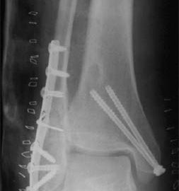
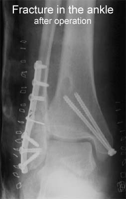
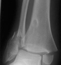
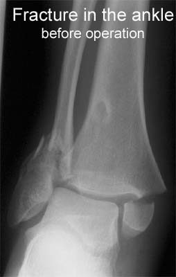
Disorders of vascularization following fracture of the ankle joint in children

Benz G, Schmid H, Daum R. Z Kinderchir 1988 Jun;43(3):183-5

In the Department of Paediatric Surgery of the University of the Hidelberg, 64 children were admitted during five years to the hospital after an ankle fracture. 12 children showed a typical fracture of the growth plate (v. Laer). All 12 were not older than 12 years. The problem of this typical form of fracture is that even careful accurate treatment may be followed by growth problems of the tibial malleolus. A microangiographic postmortem study using plastinated shetts (v. Hagens) showed a normal distribution of vascularity of the ankle joint. The vascular damage after fracture and the damage of the local vascularity in the area at the growth plate after surgical treatment are comparable.

treatment-article1

SportNetDoc

Treatment strategies for acute fractures and nonunions of the proximal fifth metatarsal.

Rosenberg GA, Sferra JJ. J Am Acad Orthop Surg 2000 Sep-Oct;8(5):332-8.

There are at least three distinct fracture patterns that occur in the proximal fifth metatarsal: tuberosity avulsion fractures, acute Jones fractures, and diaphyseal stress fractures. Each of these fracture patterns has its own mechanism of injury, location, treatment options, and prognosis regarding delayed union and nonunion. Tuberosity avulsion fractures are the most common in this region of the foot. The majority heal with symptomatic care in a hard-soled shoe. The true Jones fracture is an acute injury involving the fourth-fifth intermetatarsal facet. These injuries are best treated with non-weight-bearing cast immobilization for 6 to 8 weeks. The rate of successful union with this treatment has been reported to be between 72% and 93%. For the high-performance athlete with an acute Jones fracture, early intramedullary-screw fixation is an accepted treatment option. Nonacute diaphyseal stress fractures of the proximal fifth metatarsal and Jones fractures that develop into delayed unions and nonunions can both be managed with operative fixation with either closed axial intramedullary-screw fixation or autogenous corticocancellous grafting. Early results with the use of electrical stimulation are promising; however, prospective studies are needed to better define the role of this modality in managing these injuries.

examination-article2

SportNetDoc

Sensitivity of a clinical examination to predict need for radiography in children with ankle injuries: a prospective study.

Boutis K, Komar L, Jaramillo D, Babyn P, Alman B, Snyder B, Mandl KD, Schuh S. Lancet 2001 Dec 22-29;358(9299):2118-21

BACKGROUND: Radiographs are ordered routinely for children with ankle trauma. We assessed the predictive value of a clinical examination to identify a predefined group of low-risk injuries, management of which would not be affected by absence of a radiograph. We aimed to show that no more than 1% of children with low-risk examinations (signs restricted to the distal fibula) would have high-risk fractures (all fractures except avulsion, buckle, and non-displaced Salter-Harris I and II fractures of the distal fibula), and to compare the potential reduction in radiography in children with low-risk examinations with that obtained by application of the Ottawa ankle rules (OAR). METHODS: Standard clinical examinations and subsequent radiographs were prospectively and independently evaluated in two tertiary-care paediatric emergency departments in North America. Eligible participants were healthy children aged 3-16 years with acute ankle injuries. Sample size, negative and positive predictive values, sensitivity, and specificity were calculated. McNemar’s test was used to compare differences in the potential reduction in radiographs between the low-risk examination and the OAR. FINDINGS: 607 children were enrolled; 581 (95.7%) received follow-up. None of the 381 children with low-risk examinations had a high-risk fracture (negative predictive value 100% [95% CI 99.2-100]; sensitivity 100% [93.3-100]). Radiographs could be omitted in 62.8% of children with low-risk examinations, compared with only 12.0% reduction obtained by application of the OAR (p<0.0001). INTERPRETATION: A low-risk clinical examination in children with ankle injuries identifies 100% of high-risk diagnoses and may result in greater reduction of radiographic referrals than the OAR.

examination-article1

SportNetDoc

Acute paediatric ankle trauma: MRI versus plain radiography.

Lohman M, Kivisaari A, Kallio P, Puntila J, Vehmas T, Kivisaari L. Skeletal Radiol 2001 Sep;30(9):504-11

OBJECTIVE: To evaluate the diagnosis of acute physeal ankle fractures on plain radiographs using MRI as the gold standard. METHODS: Sixty consecutive children, 29 with a clinical diagnosis of lateral ligament injury and 31 with physeal ankle fractures, were examined using both radiographs and MRI in the acute period. The imaging data were reviewed by three “masked” radiologists. The fracture diagnosis and Slater-Harris classification of radiographs were compared with findings on MRI. RESULTS: Plain radiography produced five of 28 (18%) false negative and 12 of 92 (13%) false positive fracture diagnoses compared with MRI. Six of the 12 false positive fractures were due to a misclassification of lateral ligament disruption as SH1 fractures, Altogether a difference was found in 21% of cases in either the diagnosis or the classification of the fractures according to Salter-Harris. All bone bruises in the distal tibia and fibula and 64% of bone bruises in the talus were seen in association with lateral ligament injuries. Talar bone bruises in association with fractures occurred on the same side as the malleolar fracture; talar bone bruises in association with lateral ligament disruption were seen in different locations. The errors identified on radiographs by MRI did not affect the management of the injury. CONCLUSIONS: The incidence of false negative ankle fractures in plain radiographs was small and no complex ankle fractures were missed on radiographs. The total extent of complex fractures was, however, not always obvious on radiographs. In an unselected series of relatively mild ankle injuries, we were unable to show a single case where the treatment or prognosis based on plain radiography should have been significantly altered after having done a routine MRI examination. Plain radiography is still the diagnostic cornerstone of paediatric ankle injuries.