Kategoriarkiv: Ligament injury in the ankle joint, outer ligament

tape-description


Objective: Stabilise the ankle joint (talocrural and subtalar joints) against too great a lateral twist (supination and pronation) without reducing the normal mobility of the ankle joint (plantar and dorsal flexion). With a heavy sprain the applications include “stirrups”, “ankle lock” and possibly “figure-8”, however, light sprains normally require just “stirrups”.
The tape is utilised the whole day in the beginning of the course, being gradually reduced over the following three months. The tape is finally only used in connection with particular loads where there is risk attached.

Type: ANKLE TAPE – LATERAL STABILISATION:
“STIRRUP”
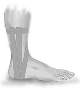
Application: The foot should be kept in a neutral position when applying the tape, at an angle of 90 degrees from the lower leg. One or two tape anchors are applied around the lower leg 10-20 cm above the ankle joint (A), followed by application of three stirrups. The stirrups are started from the anchor on the inner side of the leg if it is the outer tendons that are injured (95%), and the other way around in the rare cases where it is the inner tendons that are damaged. The stirrups are taken under the heel and drawn up to the anchor on the outer side of the leg. When applying the stirrups the outer border of the foot is pushed slightly up, whilst at the same time pulling the border slightly up with the tape. A stirrup (B) is applied with the rear half of the outer ankle knuckle directly under the tape. A sloping stirrup (D) starts from the anchor on the front side of the leg slanting down over the lower leg over the front half of the inner ankle knuckle, under the foot and over the front half of the outer ankle knuckle, and slanting in over the leg up to the anchor. Finish with a further strip on top of the anchor A.

Type: ANKLE TAPE – LATERAL STABILISATION:
“ANKLE LOCK”
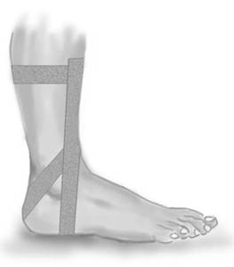
Application: One or two tape anchors are fixed around the lower leg 10-20 cm above the ankle joint (A). Begin on the anchor on the inner side of the leg, draw the tape forward in front of the leg, down over the outer ankle knuckle, behind the heel directly under the Achilles tendon, down under the heel and up over the outer side of the leg to the anchor. The ankle lock can be applied the other way around if the inner tendon is damaged. Finish with a further strip on top of the anchor A.


Type: ANKLE TAPE – LATERAL STABILISATION:
“FIGURE-8 BANDAGE”
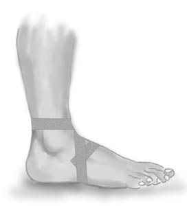
Application: Begin just in front of the ankle joint and draw the tape in under the arch of the foot on the inner side, and up over the outer side, continuing over the start tape in front of the ankle joint and round behind the leg and back again to finish in front of the ankle joint.


Type: ANKLE TAPE – LATERAL STABILISATION:
“DOUBLE FIGURE-8 BANDAGE”


Especially used with sprains of the tendon in the front of the ankle joint (lig. tibiofibular anterior), where pain is experienced when flexing the foot powerfully upwards (dorsal flexion).
This tape must be rolled loosely on, and must not be drawn tight. If the tape becomes tight all the same, which often happens, it must be loosened.

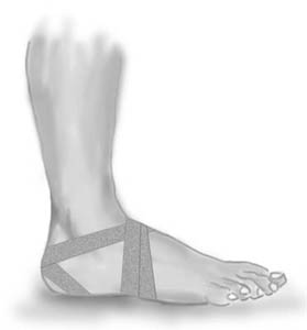
Application: Start at the back of the foot just in front of the ankle joint and draw the tape down around the outer border of the foot (A), under the sole and up to the back of the foot. Continue over the lower part of the ankle knuckle behind the heel under the Achilles tendon (B). The tape crosses down under the inner side of the heel and up to the outer border of the foot (C). The tape continues over the lower part of the inner ankle knuckle behind the heel under the Achilles tendon. The tape crosses the earlier tape, goes down under the outer side of the heel (D) and up to the inner border of the foot to finish on the back of the foot.

treatment-article2

SportNetDoc

Early functional treatment for acute ligament injuries of the ankle joint.

Karlsson J, Eriksson BI, Sward L. Scand J Med Sci Sports 1996 Dec;6(6):341-5.

Eighty-six patients with acute (< 24 h) grade II or III lateral ligament ruptures were randomized into two different non-surgical treatment groups. The main difference in treatment protocols between the two groups was during the first week after injury. Group I (n = 46) was to receive functional treatment using specially designed compression pads, elevation of the injured foot (24 h), repeated elastic wrapping (compression bandage followed by ankle tape), early full weight-bearing and proprioceptive range-of-motion training. Group II (n = 40) was to receive conventional treatment with an elastic bandage, partial weight bearing and crutches until the pain subsided. One week after the injury, patients from both groups were given identical rehabilitation instructions. No early surgery was performed. The mean follow-up period was 18 months. The functional results were satisfactory in 91% of the patients in group I and in 87% of the patients in group II (NS). Five patients had been operated on due to recurrent instability, two in group I and three in group II (NS). The mean sick leave was significantly shorter in group I; 5.6 +/- 4.2 days compared with 10.2 +/- 6.8 days in group II. Return to sports activities was also significantly earlier in group I, 9.6 +/- 4.8 days compared with 19.2 +/- 9.5 days in group II. In this study, non-surgical treatment of acute grade II and III ligament injuries of the ankle produced satisfactory results in the majority of patients. Early functional treatment resulted in shorter sick leave and facilitated an earlier return to sports, but it did not influence the final results.

treatment-article1

SportNetDoc

Treatment of ruptures of the lateral ankle ligaments: a meta-analysis.

Pijnenburg AC, Van Dijk CN, Bossuyt PM, Marti RK. J Bone Joint Surg Am 2000 Jun;82(6):761-73.

BACKGROUND.
Ruptures of the lateral ankle ligaments are very common; however, treatment remains controversial. The aim of the current study was to perform a meta-analysis of randomized, controlled clinical trials of existing treatment strategies for acute ruptures of the lateral ankle ligaments.

METHODS.
Randomized, controlled trials reported between 1966 and 1998 were included if they involved acute ruptures of the lateral ankle ligaments. Randomized, controlled trials are defined as comparative studies with an intervention group and a control group in which the assignment of participants to a group is determined by the formal procedure of randomization. Summary measures of effectiveness were expressed as relative risks with use of random effects modeling.

RESULTS.
When analyzing the trials, we searched for comparable outcome measures in both short and long-term follow-up studies (studies with six months to 3.8 years of follow-up). This resulted in the analyses of three outcome measures: time lost from work, residual pain, and giving-way. This report summarizes the results of twenty-seven trials. With respect to giving-way, a significant difference was noted between operative treatment and functional treatment (relative risk, 0.23; 95 percent confidence interval, 0.17 to 0.31) in favor of operative treatment and a significant difference was also noted between functional treatment and treatment with a cast for six weeks (relative risk, 0.69; 95 percent confidence interval, 0.50 to 0.94) in favor of functional treatment. With respect to residual pain, no significant difference was found between operative and functional treatment and a significant difference was found between functional treatment and treatment with a cast for six weeks (relative risk, 0.67; 95 percent confidence interval, 0.50 to 0.90). We found minimal or no treatment to result in more residual pain (relative risk, 0.53; 95 percent confidence interval, 0.27 to 1.02) and giving-way (relative risk, 0.34; 95 percent confidence interval, 0.17 to 0.71) than did functional treatment.

CONCLUSIONS.
We concluded that a no-treatment strategy for ruptures of the lateral ankle ligaments leads to more residual symptoms. Operative treatment leads to better results than functional treatment, and functional treatment leads to better results than cast immobilization for six weeks.

examination-article3

SportNetDoc

Is stress radiography necessary in the diagnosis of acute or chronic ankle instability?

Frost SC, Amendola A. Clin J Sport Med 1999 Jan;9(1):40-5.

BACKGROUND.
Clinicians often use the talar tilt (TT) and anterior drawer (AD) stress x-rays to diagnose acute or chronic mechanical ankle instability. However, the wide range of TT and AD values in normal and injured ankles makes interpretation of the test results difficult.

OBJECTIVE.
To critically review the literature and determine the accuracy of stress radiography in the diagnosis of mechanical ankle instability.

DATA SOURCES.
MEDLINE was searched for relevant articles published since 1966 using MEDLINE subject headings (MeSH) and textwords for English articles related to ankle injuries and radiography. Additional references were reviewed from the bibliographies of the retrieved articles. The total number of articles reviewed was 67. Of these, 8 studies met criteria for inclusion and were analyzed.

STUDY SELECTION.
Only clinical studies that used surgical exploration as the gold standard for diagnosing lateral ligament rupture were evaluated for this study. Cadaver or laboratory studies were excluded.

DATA EXTRACTION AND SYNTHESIS.
In reviewing the literature, pertinent strengths of the different study designs were emphasized. From these data, particular attention was paid to the diagnostic accuracy of each study in comparing TT and AD stress x-rays to surgical confirmation of lateral ligament rupture.

MAIN RESULTS.
A total of eight prospective clinical series satisfied the inclusion criteria. Seven of the eight assessed acute ankle instability as the outcome and one assessed chronic ankle instability. Of the seven studies that focused on acute ankle injuries, only one concluded significant benefit in using stress views to diagnose lateral ligament rupture. Three of the seven reported a positive relationship between stress radiography and surgical findings, although all six studies concluded that TT and AD stress x-rays are not reliable enough to make the diagnosis. The authors who assessed chronic ankle instability stated that TT and AD stress views combined were not useful in defining ankle instability.

CONCLUSION.
The published data regarding TT and AD stress x-rays are too variable to determine accepted normal values compared with injured values. There are insufficient data for comparison of the use of mechanical versus manual techniques, or use of local anesthetic to facilitate the stress test. Because the treatment evolution of all acute ankle sprains is toward functional nonoperative treatment and because treatment does not depend on the degree of ankle instability on stress views, the TT and AD stress x-rays have no clinical relevance in the acute situation. In cases of chronic instability, the large variability in TT and AD values in both injured and noninjured ankles precludes their routine use.

examination-article2

SportNetDoc

Sensitivity of a clinical examination to predict need for radiography in children with ankle injuries: a prospective study.

Boutis K, Komar L, Jaramillo D, Babyn P, Alman B, Snyder B, Mandl KD, Schuh S. Lancet 2001 Dec 22-29;358(9299):2118-21

BACKGROUND: Radiographs are ordered routinely for children with ankle trauma. We assessed the predictive value of a clinical examination to identify a predefined group of low-risk injuries, management of which would not be affected by absence of a radiograph. We aimed to show that no more than 1% of children with low-risk examinations (signs restricted to the distal fibula) would have high-risk fractures (all fractures except avulsion, buckle, and non-displaced Salter-Harris I and II fractures of the distal fibula), and to compare the potential reduction in radiography in children with low-risk examinations with that obtained by application of the Ottawa ankle rules (OAR). METHODS: Standard clinical examinations and subsequent radiographs were prospectively and independently evaluated in two tertiary-care paediatric emergency departments in North America. Eligible participants were healthy children aged 3-16 years with acute ankle injuries. Sample size, negative and positive predictive values, sensitivity, and specificity were calculated. McNemar’s test was used to compare differences in the potential reduction in radiographs between the low-risk examination and the OAR. FINDINGS: 607 children were enrolled; 581 (95.7%) received follow-up. None of the 381 children with low-risk examinations had a high-risk fracture (negative predictive value 100% [95% CI 99.2-100]; sensitivity 100% [93.3-100]). Radiographs could be omitted in 62.8% of children with low-risk examinations, compared with only 12.0% reduction obtained by application of the OAR (p<0.0001). INTERPRETATION: A low-risk clinical examination in children with ankle injuries identifies 100% of high-risk diagnoses and may result in greater reduction of radiographic referrals than the OAR.

cause-article2

SportNetDoc

Tendon sheath injuries of the foot and ankle.

Duddy RK, Meredith R, Visser HJ, Brooks JS. J Foot Surg 1991 Mar-Apr;30(2):179-86.

Tendon sheath injuries of the foot and ankle are a common clinical entity secondary to trauma and abnormal biomechanics. These injuries are often misdiagnosed and/or inappropriately treated. This article presents an historic review, etiology, classification, diagnosis, and treatment protocol for these injuries.

examination-article

SportNetDoc

Acute paediatric ankle trauma: MRI versus plain radiography.

Lohman M, Kivisaari A, Kallio P, Puntila J, Vehmas T, Kivisaari L. Skeletal Radiol 2001 Sep;30(9):504-11

OBJECTIVE: To evaluate the diagnosis of acute physeal ankle fractures on plain radiographs using MRI as the gold standard. METHODS: Sixty consecutive children, 29 with a clinical diagnosis of lateral ligament injury and 31 with physeal ankle fractures, were examined using both radiographs and MRI in the acute period. The imaging data were reviewed by three “masked” radiologists. The fracture diagnosis and Slater-Harris classification of radiographs were compared with findings on MRI. RESULTS: Plain radiography produced five of 28 (18%) false negative and 12 of 92 (13%) false positive fracture diagnoses compared with MRI. Six of the 12 false positive fractures were due to a misclassification of lateral ligament disruption as SH1 fractures, Altogether a difference was found in 21% of cases in either the diagnosis or the classification of the fractures according to Salter-Harris. All bone bruises in the distal tibia and fibula and 64% of bone bruises in the talus were seen in association with lateral ligament injuries. Talar bone bruises in association with fractures occurred on the same side as the malleolar fracture; talar bone bruises in association with lateral ligament disruption were seen in different locations. The errors identified on radiographs by MRI did not affect the management of the injury. CONCLUSIONS: The incidence of false negative ankle fractures in plain radiographs was small and no complex ankle fractures were missed on radiographs. The total extent of complex fractures was, however, not always obvious on radiographs. In an unselected series of relatively mild ankle injuries, we were unable to show a single case where the treatment or prognosis based on plain radiography should have been significantly altered after having done a routine MRI examination. Plain radiography is still the diagnostic cornerstone of paediatric ankle injuries.

cause-article1

SportNetDoc

Associated injuries found in chronic lateral ankle instability.

DIGiovanni BF, Fraga CJ, Cohen BE, Shereff MJ. Foot Ankle Int 2000 Oct;21(10):809-15.

Sixty-one patients underwent a primary ankle lateral ligament reconstruction for chronic instability between 1989 and 1996. In addition to the ligament reconstruction, all patients had evaluation of the peroneal retinaculum, peroneal tendon inspection by routine opening of the tendon sheath, and ankle joint inspection by arthrotomy. A retrospective review of the clinical history, physical exam, MRI examination, and intraoperative findings was conducted on these 61 patients. The purpose was to determine the type and frequency of associated injuries found at surgery and during the preoperative evaluation. At surgery no patients were found to have isolated lateral ligament injury. Fifteen different associated injuries were noted. The injuries found most often by direct inspection included: peroneal tenosynovitis, 47/61 patients (77%); anterolateral impingement lesion, 41/61 (67%); attenuated peroneal retinaculum, 33/61 (54%); and ankle synovitis, 30/61 (49%). Other less common but significant associated injuries included: intra-articular loose body, 16/61 (26%); peroneus brevis tear, 15/61 (25%); talus osteochondral lesion, 14/61 (23%); medial ankle tendon tenosynovitis, 3/61 (5%). The findings of this study indicate there is a high frequency of associated injuries in patients with chronic lateral ankle instability. Peroneal tendon and retinacular pathology, as well as anterolateral impingement lesions, occur most often. A high index of suspicion for possible associated injuries may result in more consistent outcomes with nonoperative and operative treatment of patients with chronic lateral ankle instability.