Kategoriarkiv: Foot

examination-article3

SportNetDoc

Is stress radiography necessary in the diagnosis of acute or chronic ankle instability?

Frost SC, Amendola A. Clin J Sport Med 1999 Jan;9(1):40-5.

BACKGROUND.
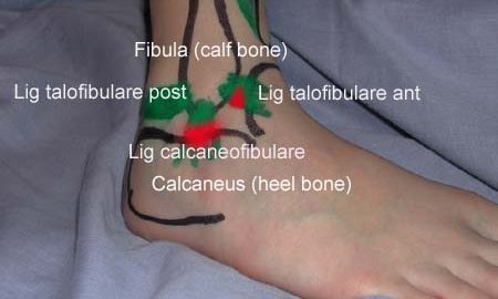
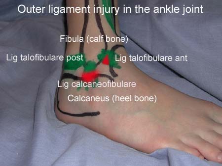
Clinicians often use the talar tilt (TT) and anterior drawer (AD) stress x-rays to diagnose acute or chronic mechanical ankle instability. However, the wide range of TT and AD values in normal and injured ankles makes interpretation of the test results difficult.

OBJECTIVE.
To critically review the literature and determine the accuracy of stress radiography in the diagnosis of mechanical ankle instability.

DATA SOURCES.
MEDLINE was searched for relevant articles published since 1966 using MEDLINE subject headings (MeSH) and textwords for English articles related to ankle injuries and radiography. Additional references were reviewed from the bibliographies of the retrieved articles. The total number of articles reviewed was 67. Of these, 8 studies met criteria for inclusion and were analyzed.

STUDY SELECTION.
Only clinical studies that used surgical exploration as the gold standard for diagnosing lateral ligament rupture were evaluated for this study. Cadaver or laboratory studies were excluded.

DATA EXTRACTION AND SYNTHESIS.
In reviewing the literature, pertinent strengths of the different study designs were emphasized. From these data, particular attention was paid to the diagnostic accuracy of each study in comparing TT and AD stress x-rays to surgical confirmation of lateral ligament rupture.

MAIN RESULTS.
A total of eight prospective clinical series satisfied the inclusion criteria. Seven of the eight assessed acute ankle instability as the outcome and one assessed chronic ankle instability. Of the seven studies that focused on acute ankle injuries, only one concluded significant benefit in using stress views to diagnose lateral ligament rupture. Three of the seven reported a positive relationship between stress radiography and surgical findings, although all six studies concluded that TT and AD stress x-rays are not reliable enough to make the diagnosis. The authors who assessed chronic ankle instability stated that TT and AD stress views combined were not useful in defining ankle instability.

CONCLUSION.
The published data regarding TT and AD stress x-rays are too variable to determine accepted normal values compared with injured values. There are insufficient data for comparison of the use of mechanical versus manual techniques, or use of local anesthetic to facilitate the stress test. Because the treatment evolution of all acute ankle sprains is toward functional nonoperative treatment and because treatment does not depend on the degree of ankle instability on stress views, the TT and AD stress x-rays have no clinical relevance in the acute situation. In cases of chronic instability, the large variability in TT and AD values in both injured and noninjured ankles precludes their routine use.

examination-article2

SportNetDoc

Sensitivity of a clinical examination to predict need for radiography in children with ankle injuries: a prospective study.

Boutis K, Komar L, Jaramillo D, Babyn P, Alman B, Snyder B, Mandl KD, Schuh S. Lancet 2001 Dec 22-29;358(9299):2118-21

BACKGROUND: Radiographs are ordered routinely for children with ankle trauma. We assessed the predictive value of a clinical examination to identify a predefined group of low-risk injuries, management of which would not be affected by absence of a radiograph. We aimed to show that no more than 1% of children with low-risk examinations (signs restricted to the distal fibula) would have high-risk fractures (all fractures except avulsion, buckle, and non-displaced Salter-Harris I and II fractures of the distal fibula), and to compare the potential reduction in radiography in children with low-risk examinations with that obtained by application of the Ottawa ankle rules (OAR). METHODS: Standard clinical examinations and subsequent radiographs were prospectively and independently evaluated in two tertiary-care paediatric emergency departments in North America. Eligible participants were healthy children aged 3-16 years with acute ankle injuries. Sample size, negative and positive predictive values, sensitivity, and specificity were calculated. McNemar’s test was used to compare differences in the potential reduction in radiographs between the low-risk examination and the OAR. FINDINGS: 607 children were enrolled; 581 (95.7%) received follow-up. None of the 381 children with low-risk examinations had a high-risk fracture (negative predictive value 100% [95% CI 99.2-100]; sensitivity 100% [93.3-100]). Radiographs could be omitted in 62.8% of children with low-risk examinations, compared with only 12.0% reduction obtained by application of the OAR (p<0.0001). INTERPRETATION: A low-risk clinical examination in children with ankle injuries identifies 100% of high-risk diagnoses and may result in greater reduction of radiographic referrals than the OAR.

cause-article2

SportNetDoc

Tendon sheath injuries of the foot and ankle.

Duddy RK, Meredith R, Visser HJ, Brooks JS. J Foot Surg 1991 Mar-Apr;30(2):179-86.

Tendon sheath injuries of the foot and ankle are a common clinical entity secondary to trauma and abnormal biomechanics. These injuries are often misdiagnosed and/or inappropriately treated. This article presents an historic review, etiology, classification, diagnosis, and treatment protocol for these injuries.

examination-article

SportNetDoc

Acute paediatric ankle trauma: MRI versus plain radiography.

Lohman M, Kivisaari A, Kallio P, Puntila J, Vehmas T, Kivisaari L. Skeletal Radiol 2001 Sep;30(9):504-11

OBJECTIVE: To evaluate the diagnosis of acute physeal ankle fractures on plain radiographs using MRI as the gold standard. METHODS: Sixty consecutive children, 29 with a clinical diagnosis of lateral ligament injury and 31 with physeal ankle fractures, were examined using both radiographs and MRI in the acute period. The imaging data were reviewed by three “masked” radiologists. The fracture diagnosis and Slater-Harris classification of radiographs were compared with findings on MRI. RESULTS: Plain radiography produced five of 28 (18%) false negative and 12 of 92 (13%) false positive fracture diagnoses compared with MRI. Six of the 12 false positive fractures were due to a misclassification of lateral ligament disruption as SH1 fractures, Altogether a difference was found in 21% of cases in either the diagnosis or the classification of the fractures according to Salter-Harris. All bone bruises in the distal tibia and fibula and 64% of bone bruises in the talus were seen in association with lateral ligament injuries. Talar bone bruises in association with fractures occurred on the same side as the malleolar fracture; talar bone bruises in association with lateral ligament disruption were seen in different locations. The errors identified on radiographs by MRI did not affect the management of the injury. CONCLUSIONS: The incidence of false negative ankle fractures in plain radiographs was small and no complex ankle fractures were missed on radiographs. The total extent of complex fractures was, however, not always obvious on radiographs. In an unselected series of relatively mild ankle injuries, we were unable to show a single case where the treatment or prognosis based on plain radiography should have been significantly altered after having done a routine MRI examination. Plain radiography is still the diagnostic cornerstone of paediatric ankle injuries.

cause-article1

SportNetDoc

Associated injuries found in chronic lateral ankle instability.

DIGiovanni BF, Fraga CJ, Cohen BE, Shereff MJ. Foot Ankle Int 2000 Oct;21(10):809-15.

Sixty-one patients underwent a primary ankle lateral ligament reconstruction for chronic instability between 1989 and 1996. In addition to the ligament reconstruction, all patients had evaluation of the peroneal retinaculum, peroneal tendon inspection by routine opening of the tendon sheath, and ankle joint inspection by arthrotomy. A retrospective review of the clinical history, physical exam, MRI examination, and intraoperative findings was conducted on these 61 patients. The purpose was to determine the type and frequency of associated injuries found at surgery and during the preoperative evaluation. At surgery no patients were found to have isolated lateral ligament injury. Fifteen different associated injuries were noted. The injuries found most often by direct inspection included: peroneal tenosynovitis, 47/61 patients (77%); anterolateral impingement lesion, 41/61 (67%); attenuated peroneal retinaculum, 33/61 (54%); and ankle synovitis, 30/61 (49%). Other less common but significant associated injuries included: intra-articular loose body, 16/61 (26%); peroneus brevis tear, 15/61 (25%); talus osteochondral lesion, 14/61 (23%); medial ankle tendon tenosynovitis, 3/61 (5%). The findings of this study indicate there is a high frequency of associated injuries in patients with chronic lateral ankle instability. Peroneal tendon and retinacular pathology, as well as anterolateral impingement lesions, occur most often. A high index of suspicion for possible associated injuries may result in more consistent outcomes with nonoperative and operative treatment of patients with chronic lateral ankle instability.

complication-article

SportNetDoc

Disorders of vascularization following fracture of the ankle joint in children

Benz G, Schmid H, Daum R. Z Kinderchir 1988 Jun;43(3):183-5

In the Department of Paediatric Surgery of the University of the Hidelberg, 64 children were admitted during five years to the hospital after an ankle fracture. 12 children showed a typical fracture of the growth plate (v. Laer). All 12 were not older than 12 years. The problem of this typical form of fracture is that even careful accurate treatment may be followed by growth problems of the tibial malleolus. A microangiographic postmortem study using plastinated shetts (v. Hagens) showed a normal distribution of vascularity of the ankle joint. The vascular damage after fracture and the damage of the local vascularity in the area at the growth plate after surgical treatment are comparable.

treatment-article

SportNetDoc

Osteochondral injuries. Clinical findings.

Birk GT, DeLee JC. Clin Sports Med 2001 Apr;20(2):279-86

Osteochondral injuries are common, mainly affecting a young, active population. Failure to recognize these injuries can lead to long-term disability. A heightened awareness and understanding of the common mechanisms of injury will lead to early diagnosis and rapid recovery. This, along with improved treatment modalities, will keep any long-term disability to a minimum.