|
Treatment strategies in osteochondral defects of the talar dome: a systematic review. |
Kategoriarkiv: Foot
treatment-article1
|
Arthroscopic management of osteochondral lesions of the talus: results of drilling and usefulness of magnetic resonance imaging before and after treatment. |
examination-article
|
The staging of osteochondritis dissecans in the knee and ankle joints with MR tomography. A comparison with conventional radiology and arthroscopy. |
treatment-article1
|
Treatment strategies for acute fractures and nonunions of the proximal fifth metatarsal. |
examination-article2
|
Sensitivity of a clinical examination to predict need for radiography in children with ankle injuries: a prospective study. |
examination-article1
|
Acute paediatric ankle trauma: MRI versus plain radiography. |
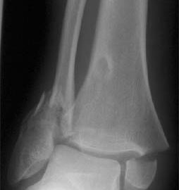
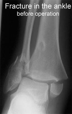
Cause(x-ray picture)
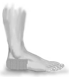
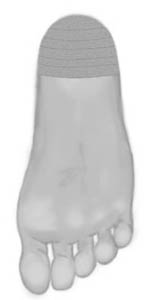
tape-description
Objective: Gather the heel pad under the heel to improve shock absorption. |
|||
  |
|||
| Application: Apply a semicircle around the heel (A) – “anchor”. The heel pad is gathered under the heel with several strips of tape (B). No bare skin should be visible. Finish with a further strip on top of the “anchor” A. |
Symptoms
|
Black heel a minor hazard of sport. |