Kategoriarkiv: Knee
STEP4
TRAINING LADDER FOR CHILDREN AND ADOLESCENTS:
FOR LUXATION OF THE KNEE CAP
(LUXATIO PATELLAE)
STEP 4 |
Unlimited: Cycling. Swimming. Running and spurting with sudden directional change.
|
||||||||||||||||||||||||||||||||||||||||||||||
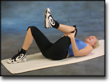
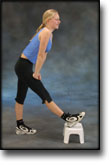
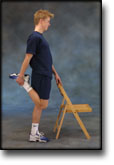
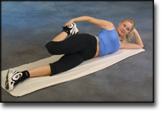
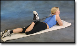
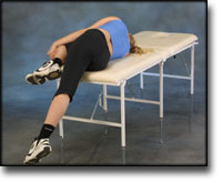
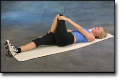
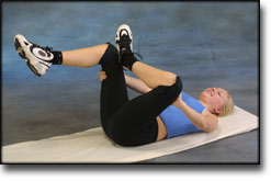
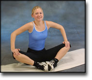
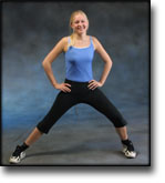


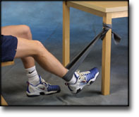
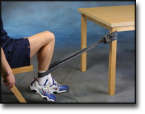
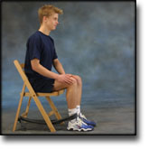
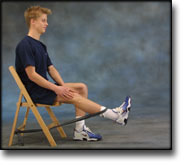
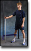
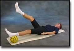
| Stretching is carried out in the following way: stretch the muscle group for 3-5 seconds. Relax for 3-5 seconds. The muscle group should subsequently be stretched for 20 seconds. The muscle is allowed to be tender, but must not hurt. Relax for 20 seconds, after which the procedure can be repeated.
The time consumed for stretching, coordination and strength training can be altered depending on the training opportunities available and individual requirements. |
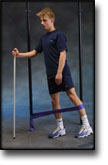
STEP3
TRAINING LADDER FOR CHILDREN AND ADOLESCENTS:
FOR LUXATION OF THE KNEE CAP
(LUXATIO PATELLAE)
STEP 3 |
Unlimited: Cycling. Swimming. Running with increasing speed and cautious directional change.
|
||||||||||||||||||||||||||||||||
| Stretching is carried out in the following way: stretch the muscle group for 3-5 seconds. Relax for 3-5 seconds. The muscle group should subsequently be stretched for 20 seconds. The muscle is allowed to be tender, but must not hurt. Relax for 20 seconds, after which the procedure can be repeated.
The time consumed for stretching, coordination and strength training can be altered depending on the training opportunities available and individual requirements. |
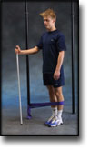
STEP2
TRAINING LADDER FOR CHILDREN AND ADOLESCENTS:
FOR LUXATION OF THE KNEE CAP
(LUXATIO PATELLAE)
STEP 2 |
Unlimited: Cycling. Swimming. Light jogging.
|
||||||||||||||||||||||||||||||||
| Stretching is carried out in the following way: stretch the muscle group for 3-5 seconds. Relax for 3-5 seconds. The muscle group should subsequently be stretched for 20 seconds. The muscle is allowed to be tender, but must not hurt. Relax for 20 seconds, after which the procedure can be repeated.
The time consumed for stretching, coordination and strength training can be altered depending on the training opportunities available and individual requirements. |
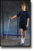
STEP1
TRAINING LADDER FOR CHILDREN AND ADOLESCENTS:
FOR LUXATION OF THE KNEE CAP
(LUXATIO PATELLAE)
STEP 1 |
Unlimited: Cycling with raised saddle. Swimming (crawl).
|
||||||||||||||||||||||||||||||
| Stretching is carried out in the following way: stretch the muscle group for 3-5 seconds. Relax for 3-5 seconds. The muscle group should subsequently be stretched for 20 seconds. The muscle is allowed to be tender, but must not hurt. Relax for 20 seconds, after which the procedure can be repeated.
The time consumed for stretching, coordination and strength training can be altered depending on the training opportunities available and individual requirements. |
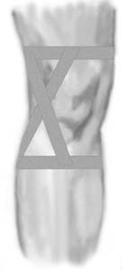
tape-description
Objective: To control the movement of the kneecap so that it glides up and down in a direct line. The tape should immediately provide less pain, and is a good indicator of whether the patient will gain any benefit of a kneecap stabilising bandage. |
|||
 |
|||
| Application: The knee should be held slightly bent when applying the tape. One anchor is applied above the knee and one below (A & B), and should be open to the rear. A strip is applied (C) over the kneecap’s lower outer half, so that half the tape is over the kneecap. The tape is in line with the edge of the kneecap, and in this way has a sloping course, lifting and pushing the kneecap slightly inwards. The second strip (D) is applied over the kneecap’s upper outer half, so that half the tape is over the kneecap and runs slanting downwards over the first strip. Finish with further strips on top of the anchors A & B. |
treatment-article2
|
Femoropatellar pain syndrome. Conservative treatment and results 7-10 years following Maquet operation. |
treatment-article1
|
Chondromalacia of the patella. |
examination-article1
|
The initial (I and II) and advanced (III and IV) stages of juvenile patellar chondromalacia. Its diagnosis by magnetic resonance using a 1.5-T magnet with FLASH sequences.
|
cause-article1
|
Anterior knee pain syndrome: a historical review. |