Kategoriarkiv: Meniscus lesion

KONDITION

STEP4

Training ladder for:
MENISCUS LESION
(LAESIO TRAUMATICA MENISCI)

STEP 4

Training must not bring about swelling in the knee.
KONDITION
Unlimited: Cycling. Swimming. Running and spurting with sudden directional change and jumping.

UDSPÆNDING
(5 min)

Lie on your back. Draw the injured leg up towards your head so that the muscles in the back of the thigh become increasingly stretched. Perform the exercise with outstretched as well as bent knee. Hold the position for 20 seconds and relax for 20 seconds before repeating. The exercise can also be performed standing with the injured leg outstretched on a chair while the upper body is bent slightly forwards.

Stand with support from the back of a chair or the wall. Using your hand, bend the knee and draw the foot up and your knee slightly backwards so that the muscles in the front of the thigh become increasingly stretched. Hold the position for 20 seconds and relax for 20 seconds before repeating. The exercise can also be done lying down. If you lie on your stomach you can draw the foot up by using a towel.

KOORDINATION
(5 min)

Seesaw. Balance on two legs, possibly using a hand as support against the wall, balancing subsequently on one leg without support. Look straight ahead and keep knees bent.

STYRKE
(10 min)

Sit on a chair with elastic around the ankle, facing the elastic. Lift the leg and slowly bend and stretch the knee.

Sit on a chair with elastic attached to the ankle. Raise the leg and slowly stretch and bend the knee.

Stand on the healthy leg with the elastic around the inside of the injured leg. Move the injured leg from side to side in a slow smooth movement. Moving the position of the elastic lower down the leg can increase the load.

Stand on the healthy leg with the elastic around the outside of the injured leg. Move the injured leg from side to side in a slow smooth movement. Moving the position of the elastic lower down the leg can increase the load.

Stand with your back against a wall with a ball or firm round cushion between the wall and your back. Slowly go down to bend your knee 90 degrees before slowly rising up again.

Stretching is carried out in the following way: stretch the muscle group for 3-5 seconds. Relax for 3-5 seconds. The muscle group should subsequently be stretched for 20 seconds. The muscle is allowed to be tender, but must not hurt. Relax for 20 seconds, after which the procedure can be repeated.

The time consumed for stretching, coordination and strength training can be altered depending on the training opportunities available and individual requirements.

KONDITION

STEP3

Training ladder for:
MENISCUS LESION
(LAESIO TRAUMATICA MENISCI)

STEP 3

Training must not bring about swelling in the knee.
KONDITION
Unlimited: Cycling. Swimming. Running with increasing speed and cautious directional change.

UDSPÆNDING
(5 min)

Lie on your back. Draw the injured leg up towards your head so that the muscles in the back of the thigh become increasingly stretched. Perform the exercise with outstretched as well as bent knee. Hold the position for 20 seconds and relax for 20 seconds before repeating. The exercise can also be performed standing with the injured leg outstretched on a chair while the upper body is bent slightly forwards.

Stand with support from the back of a chair or the wall. Using your hand, bend the knee and draw the foot up and your knee slightly backwards so that the muscles in the front of the thigh become increasingly stretched. Hold the position for 20 seconds and relax for 20 seconds before repeating. The exercise can also be done lying down. If you lie on your stomach you can draw the foot up by using a towel.

KOORDINATION
(5 min)

Seesaw. Balance on two legs, possibly using a hand as support against the wall, balancing subsequently on one leg without support. Look straight ahead and keep knees bent.

STYRKE
(10 min)

Sit on a chair with elastic around the ankle, facing the elastic. Lift the leg and slowly bend and stretch the knee.

Sit on a chair with elastic attached to the ankle. Raise the leg and slowly stretch and bend the knee.

Stand on the healthy leg with the elastic around the inside of the injured leg. Move the injured leg from side to side in a slow smooth movement. Moving the position of the elastic lower down the leg can increase the load.

Stand on the healthy leg with the elastic around the outside of the injured leg. Move the injured leg from side to side in a slow smooth movement. Moving the position of the elastic lower down the leg can increase the load.

Stand with your back to the wall with your weight on both feet. Slowly go down and bend the knee to 90 degrees, and slowly rise again.

Stretching is carried out in the following way: stretch the muscle group for 3-5 seconds. Relax for 3-5 seconds. The muscle group should subsequently be stretched for 20 seconds. The muscle is allowed to be tender, but must not hurt. Relax for 20 seconds, after which the procedure can be repeated.

The time consumed for stretching, coordination and strength training can be altered depending on the training opportunities available and individual requirements.

KONDITION

STEP2

Training ladder for:
MENISCUS LESION
(LAESIO TRAUMATICA MENISCI)

STEP 2

Training must not bring about swelling in the knee.
KONDITION
Unlimited: Cycling. Swimming. Light jogging.

UDSPÆNDING
(5 min)

Lie on your back. Draw the injured leg up towards your head so that the muscles in the back of the thigh become increasingly stretched. Perform the exercise with outstretched as well as bent knee. Hold the position for 20 seconds and relax for 20 seconds before repeating. The exercise can also be performed standing with the injured leg outstretched on a chair while the upper body is bent slightly forwards.

Stand with support from the back of a chair or the wall. Using your hand, bend the knee and draw the foot up and your knee slightly backwards so that the muscles in the front of the thigh become increasingly stretched. Hold the position for 20 seconds and relax for 20 seconds before repeating. The exercise can also be done lying down. If you lie on your stomach you can draw the foot up by using a towel.

KOORDINATION
(5 min)

Seesaw. Balance on two legs, possibly using a hand as support against the wall, balancing subsequently on one leg without support. Look straight ahead and keep knees bent.

STYRKE
(10 min)

Sit on a chair with elastic around the ankle, facing the elastic. Lift the leg and slowly bend and stretch the knee.

Sit on a chair with elastic attached to the ankle. Raise the leg and slowly stretch and bend the knee.

Stand on the healthy leg with the elastic around the inside of the injured leg. Move the injured leg from side to side in a slow smooth movement. Moving the position of the elastic lower down the leg can increase the load.

Stand on the healthy leg with the elastic around the outside of the injured leg. Move the injured leg from side to side in a slow smooth movement. Moving the position of the elastic lower down the leg can increase the load.

Stretching is carried out in the following way: stretch the muscle group for 3-5 seconds. Relax for 3-5 seconds. The muscle group should subsequently be stretched for 20 seconds. The muscle is allowed to be tender, but must not hurt. Relax for 20 seconds, after which the procedure can be repeated.

The time consumed for stretching, coordination and strength training can be altered depending on the training opportunities available and individual requirements.

KONDITION

STEP1

Training ladder for:
MENISCUS LESION
(LAESIO TRAUMATICA MENISCI)

STEP 1

Training must not bring about swelling in the knee.
KONDITION
Unlimited: Cycling with raised saddle.

KOORDINATION
(5 min)

Seesaw. Balance on two legs, possibly using a hand as support against the wall, balancing subsequently on one leg without support. Look straight ahead and keep knees bent.

Stretching is carried out in the following way: stretch the muscle group for 3-5 seconds. Relax for 3-5 seconds. The muscle group should subsequently be stretched for 20 seconds. The muscle is allowed to be tender, but must not hurt. Relax for 20 seconds, after which the procedure can be repeated.

The time consumed for stretching, coordination and strength training can be altered depending on the training opportunities available and individual requirements.

treatment-article2

SportNetDoc

Meniscal injury in children: long-term results after meniscectomy.

Dai L, Zhang W, Xu Y. Knee Surg Sports Traumatol Arthrosc 1997;5(2):77-9

Twenty-four children who underwent open total meniscectomy were reviewed and followed-up for an average of 16.1 years (range 6-33 years). There were 7 boys and 17 girls aged between 7 and 16 years (average 12.5 years). Excellent and good results were noted in 62.5% of the patients. Eighty-seven of the children showed radiographic degeneration of the knee joint. This study in a Chinese population indicates that meniscectomy is not a benign procedure in children, and total meniscectomy should be avoided as far as possible.

treatment-article1

SportNetDoc

Treatment of intrasubstance meniscal lesions: a randomized prospective study of four different methods.

Biedert RM. Knee Surg Sports Traumatol Arthrosc 2000;8(2):104-8.

This study examined the effect of four different methods for treating intrasubstance meniscal lesions. Forty patients (21 men, 19 women; age 30.4 years, range 16-50) with an isolated and symptomatic painful horizontal grade 2 meniscal lesion on the medial side (documented with MRI) were included. Patients were randomly assigned by the birth date to one of four treatment groups: group A, conservative therapy (n = 12); group B, arthroscopic suture repair with access channels (n = 10); group C, arthroscopic minimal central resection, intrameniscal fibrin clot and suture repair (n = 7); and group D, arthroscopic partial meniscectomy (n = 11). The average length of follow-up was 26.5 months (range 12-38 months). Follow-up evaluation consisted of clinical examination with the findings recorded according to the IKDC protocol, radiographs, and control MRI. Group A had 75% normal or nearly normal final evaluation at follow-up, group B 90%, group C 43%, and group D 100% normal or nearly normal at follow-up. These short-term results indicate that intrasubstance meniscal lesions can be treated best by performing partial meniscectomy. To preserve the important function of the meniscus, arthroscopic suture repair with access channels might give even better medium- to long-term results. Conservative treatment is often not satisfactory. Additionally, our findings show that MRI examinations are not superior to accurate clinical examinations.

examination-article1

SportNetDoc

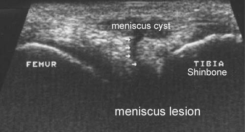
3-dimensional ultrasound in clinical diagnosis of meniscus lesions.

Riedl S, Tauscher A, Kuhner C, Gohring U, Sohn C, Meeder PJ. Ultraschall Med 1998 Feb;19(1):28-33.

AIM.
To compare the significance of the two-dimensional and three-dimensional sonography in the diagnosis of meniscal tears under clinical conditions.

METHODS.
Sixty menisci of knees with clinical symptoms (44 medial and 16 lateral menisci) were examined by an identical transducer in a two- and three-dimensional sonography technique. The findings were compared with the diagnosis made with subsequent arthroscopy.

RESULTS.
In the diagnosis of meniscal lesions the two- and three-dimensional sonography reached a sensitivity of 92% and 100% and a specificity of 83% and 88%, respectively. The positive predictive value of these methods was 58% and 67%. The negative predictive value was 98% and 100%. Because of the good results with the two-dimensional sonography, there were no statistically significant differences between both methods. The three-dimensional sonography, however, proved to be superior, to the two-dimensional sonography in the analysis of subgroups (medial and lateral menisci, menisci with and without clinical symptoms). The negative predictive value of the three-dimensional sonography was 100% for all of these subgroups.

CONCLUSION.
Although this study shows no significant difference in the results of two- and three-dimensional sonography, the analysis of subgroups displays a slightly improved significance for diagnosis of meniscal tears by three-dimensional sonography. The high negative predictive value shows that three-dimensional sonography, performed by an experienced examiner, may be useful to exclude meniscal tears. This result may help focus further cost-intensive or invasive examinations.

cause-article2

SportNetDoc

Meniscal repair in very young children.

Bloome DM, Blevins FT, Paletta GA Jr, Newcomer JK, Cashmore B, Turker R. Arthroscopy 2000 Jul-Aug;16(5):545-9.

This article reports the cases of what we believe to be the youngest patients with traumatic meniscal tears treated by repair. The 2 cases were treated with different meniscal repair techniques. A review of the literature regarding traumatic meniscal tears in children is provided.