Kategoriarkiv: Inflamed mucous fold (plica synovialis)

examination-article

SportNetDoc

The plica syndrome: diagnostic value of MRI with arthroscopic correlation.

Jee WH, Choe BY, Kim JM, Song HH, Choi KH. J Comput Assist Tomogr 1998 Sep-Oct;22(5):814-8.

PURPOSE.
Our goal was to evaluate the diagnostic value of MRI in plica syndrome.

METHOD.
MR images of a patient group (n = 55) with arthroscopically confirmed pathologic mediopatellar plicae were retrospectively analyzed and compared with those of a control group (n = 100). We obtained axial multiplanar gradient-recalled (MPGR), axial T1-weighted, and sagittal T2-weighted MR images. MR images were assessed for the width and length of all medial plicae.

RESULTS.
In the diagnosis of plica syndrome, sensitivity and specificity were 73 and 78% on axial MPGR images, 71 and 83% on sagittal T2-weighted images, and 95 and 72% on combination of both images, respectively. The incidence of pathologic medial plica increased with a criterion of extension beyond the medial end of the patella on axial MPGR images.

CONCLUSION.
MRI is a useful screening method in the diagnosis of plica syndrome.

symptoms-article

SportNetDoc

Synovial plicae of the knee. Controversies and review.

Dupont JY. Clin Sports Med 1997 Jan;16(1):87-122.

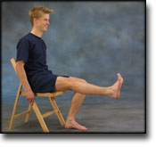
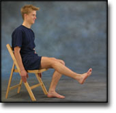
Plicae are some of the normal synovial structures of the knee joint cavity. They are remnants of the mesenchymal tissue that occupies the space between the distal femoral and proximal tibial epiphyses in the 8-week-old embryo. The incomplete resorption leaves synovial pleats in most of the knee. The superior and the inferior plicae are the most common (50% to 65%) but have extremely little clinical relevance. Each may be of many various morphological types. The lateral plica is rare (1% to 3%). The medial plica is present at autopsies in one of every three or four knees. It also is of various types, wide and thick in one of every fifteen knees. Arthrography, ultrasonography, CT scan with arthrography, and MR imaging can demonstrate their presence and measure their size with good accuracy. Arthroscopy allows a very precise assessment of the plica, including dynamic examination. It looks for medial impingement against the patellofemoral articular surfaces and secondary (localized chondromalacia) as well as incidentally associated other knee pathologic conditions. Rarely, the medial plica becomes symptomatic, circumstances such as a history of blunt trauma, or more often, overuse of the knee can cause symptoms. Sometimes no special condition is necessary. The plica causes symptoms such as pain, crepitus, snapping or popping, or effusion related to patellofemoral joint motion. The clinical picture mimics a torn medial meniscus or a maltracking patella. Clinical examination is extremely helpful if the snapping plica is palpated at the medial edge of the patella, reproducing the patient’s symptoms. If chronic, these symptoms may be treated with nonsteroidal anti-inflammatory drugs, physiotherapy, electrophoresis, or local injection. Surgical treatment is indicated if conservative therapy fails. Arthroscopic complete resection of the plica cures the symptoms in a few days, therefore confirming the correct diagnosis and the effectiveness of the treatment. Histologic examination often confirms the chronic conflict between the plica and the femoral condyle. No morphologic character allows the assessment of the pathologic aspect of the plica. A medial plica is or is not symptomatic. The incidence of this syndrome is probably one out of ten medial plicae and 3% of arthroscopies at most. Associated lesions are very common. They often make the evaluation of the plica’s responsibility in symptoms difficult to analyze, leading to unsatisfactory results.

anatomy-article1

SportNetDoc

Plica: Pathologic or Not?

Ewing JW. J Am Acad Orthop Surg 1993 Nov;1(2):117-121.

A fold that occurs within a joint is referred to as a plica synovialis. Three such plicae are seen with regularity within the human knee joint. These folds are normal structures that represent remnants of mesenchymal tissue and/or septa formed during embryonic development of the knee joint, and can be seen during arthroscopic inspection of the knee joint. Controversy exists within the orthopaedic community as to whether a plica can develop pathologic changes sufficient to cause disabling knee symptoms. The author defines the clinical syndrome, describes the arthroscopic appearance of pathologic plica, and outlines nonsurgical and surgical methods of management of this uncommon condition.

KONDITION

step4

Training ladder for:
INFLAMMATION OF THE MUCOUS FOLD
(PLICA SYNOVIALIS)

STEP 4

Training must not bring about swelling in the knee.
KONDITION
Unlimited: Cycling. Swimming. Running and spurting with sudden directional change and jumping.

UDSPÆNDING
(5 min)

Lie on your back. Draw the injured leg up towards your head so that the muscles in the back of the thigh become increasingly stretched. Perform the exercise with outstretched as well as bent knee. Hold the position for 20 seconds and relax for 20 seconds before repeating. The exercise can also be performed standing with the injured leg outstretched on a chair while the upper body is bent slightly forwards.

Stand with support from the back of a chair or the wall. Using your hand, bend the knee and draw the foot up and your knee slightly backwards so that the muscles in the front of the thigh become increasingly stretched. Hold the position for 20 seconds and relax for 20 seconds before repeating. The exercise can also be done lying down. If you lie on your stomach you can draw the foot up by using a towel.

Lie on your side on a table. Bend one leg up under your body and let the other hang over the edge of the table so that the muscles in the outer side of the thigh become increasingly stretched. Hold the position for 20 seconds and relax for 20 seconds before repeating. The exercise can also be done standing by placing the outstretched injured leg behind the good leg at the same time as bending over the injured leg.

Stand with one leg outstretched and the other slightly bent. Thrust your weight to the side over the bent leg so that the inner side of the opposite thigh becomes increasingly stretched. Hold the position for 20 seconds and relax for 20 seconds before repeating.

KOORDINATION
(5 min)

Seesaw. Balance on two legs, possibly using a hand as support against the wall, balancing subsequently on one leg without support. Look straight ahead and keep knees bent.

STYRKE
(50 min)

Sit on a chair with elastic around the ankle, facing the elastic. Lift the leg and slowly bend and stretch the knee.

Stand on the healthy leg with the elastic around the inside of the injured leg. Move the injured leg from side to side in a slow smooth movement. Moving the position of the elastic lower down the leg can increase the load.

Stand on the healthy leg with the elastic around the outside of the injured leg. Move the injured leg from side to side in a slow smooth movement. Moving the position of the elastic lower down the leg can increase the load.

Stand with elastic around the hip. Step forward over one knee and hold the front foot firmly against the floor. Bend the rear leg and go forward onto your toes. Remember to change leg.

Stand with your back against a wall with a ball or firm round cushion between the wall and your back. Slowly go down to bend your knee 90 degrees before slowly rising up again.

Lie on your back with a ball or firm round cushion under the injured leg. Lift your backside up from the floor and stretch the healthy leg. Hold the position for a few seconds.

Lie on your back with a ball or firm round cushion under both feet. Roll the ball backwards and forwards in a steady pace while lifting your backside.

Stretching is carried out in the following way: stretch the muscle group for 3-5 seconds. Relax for 3-5 seconds. The muscle group should subsequently be stretched for 20 seconds. The muscle is allowed to be tender, but must not hurt. Relax for 20 seconds, after which the procedure can be repeated. The time consumed for stretching, coordination and strength training can be altered depending on the training opportunities available and individual requirements.

KONDITION

step3

Training ladder for:
INFLAMMATION OF THE MUCOUS FOLD
(PLICA SYNOVIALIS)

STEP 3

Training must not bring about swelling in the knee.
KONDITION
Unlimited: Cycling. Swimming. Running with increasing speed and cautious directional change.

UDSPÆNDING
(5 min)

Lie on your back. Draw the injured leg up towards your head so that the muscles in the back of the thigh become increasingly stretched. Perform the exercise with outstretched as well as bent knee. Hold the position for 20 seconds and relax for 20 seconds before repeating. The exercise can also be performed standing with the injured leg outstretched on a chair while the upper body is bent slightly forwards.

Stand with support from the back of a chair or the wall. Using your hand, bend the knee and draw the foot up and your knee slightly backwards so that the muscles in the front of the thigh become increasingly stretched. Hold the position for 20 seconds and relax for 20 seconds before repeating. The exercise can also be done lying down. If you lie on your stomach you can draw the foot up by using a towel.

Lie on your side on a table. Bend one leg up under your body and let the other hang over the edge of the table so that the muscles in the outer side of the thigh become increasingly stretched. Hold the position for 20 seconds and relax for 20 seconds before repeating. The exercise can also be done standing by placing the outstretched injured leg behind the good leg at the same time as bending over the injured leg.

Stand with one leg outstretched and the other slightly bent. Thrust your weight to the side over the bent leg so that the inner side of the opposite thigh becomes increasingly stretched. Hold the position for 20 seconds and relax for 20 seconds before repeating.

KOORDINATION
(5 min)

Seesaw. Balance on two legs, possibly using a hand as support against the wall, balancing subsequently on one leg without support. Look straight ahead and keep knees bent.

STYRKE
(50 min)

Sit on a chair with elastic around the ankle, facing the elastic. Lift the leg and slowly bend and stretch the knee.

Stand on the healthy leg with the elastic around the inside of the injured leg. Move the injured leg from side to side in a slow smooth movement. Moving the position of the elastic lower down the leg can increase the load.

Stand on the healthy leg with the elastic around the outside of the injured leg. Move the injured leg from side to side in a slow smooth movement. Moving the position of the elastic lower down the leg can increase the load.

Stand with elastic around the hip. Step forward over one knee and hold the front foot firmly against the floor. Bend the rear leg and go forward onto your toes. Remember to change leg.

Stand with your back to the wall with your weight on both feet. Slowly go down and bend the knee to 90 degrees, and slowly rise again.

Lie on your back with a ball or firm round cushion under the injured leg. Lift your backside up from the floor and stretch the healthy leg. Hold the position for a few seconds.

Lie on your back with a ball or firm round cushion under both feet. Roll the ball backwards and forwards in a steady pace while lifting your backside.

Stretching is carried out in the following way: stretch the muscle group for 3-5 seconds. Relax for 3-5 seconds. The muscle group should subsequently be stretched for 20 seconds. The muscle is allowed to be tender, but must not hurt. Relax for 20 seconds, after which the procedure can be repeated. The time consumed for stretching, coordination and strength training can be altered depending on the training opportunities available and individual requirements.

KONDITION

step2

Training ladder for:
INFLAMMATION OF THE MUCOUS FOLD
(PLICA SYNOVIALIS)

STEP 2

Training must not bring about swelling in the knee.
KONDITION
Unlimited: Cycling. Swimming. Light jogging.

UDSPÆNDING
(5 min)

Lie on your back. Draw the injured leg up towards your head so that the muscles in the back of the thigh become increasingly stretched. Perform the exercise with outstretched as well as bent knee. Hold the position for 20 seconds and relax for 20 seconds before repeating. The exercise can also be performed standing with the injured leg outstretched on a chair while the upper body is bent slightly forwards.

Stand with support from the back of a chair or the wall. Using your hand, bend the knee and draw the foot up and your knee slightly backwards so that the muscles in the front of the thigh become increasingly stretched. Hold the position for 20 seconds and relax for 20 seconds before repeating. The exercise can also be done lying down. If you lie on your stomach you can draw the foot up by using a towel.

Lie on your side on a table. Bend one leg up under your body and let the other hang over the edge of the table so that the muscles in the outer side of the thigh become increasingly stretched. Hold the position for 20 seconds and relax for 20 seconds before repeating. The exercise can also be done standing by placing the outstretched injured leg behind the good leg at the same time as bending over the injured leg.

Stand with one leg outstretched and the other slightly bent. Thrust your weight to the side over the bent leg so that the inner side of the opposite thigh becomes increasingly stretched. Hold the position for 20 seconds and relax for 20 seconds before repeating.

KOORDINATION
(5 min)

Seesaw. Balance on two legs, possibly using a hand as support against the wall, balancing subsequently on one leg without support. Look straight ahead and keep knees bent.

STYRKE
(50 min)

Sit on a chair with elastic around the ankle, facing the elastic. Lift the leg and slowly bend and stretch the knee.

Sit on a chair with elastic attached to the ankle. Raise the leg and slowly stretch and bend the knee.

Stand on the healthy leg with the elastic around the inside of the injured leg. Move the injured leg from side to side in a slow smooth movement. Moving the position of the elastic lower down the leg can increase the load.

Stand on the healthy leg with the elastic around the outside of the injured leg. Move the injured leg from side to side in a slow smooth movement. Moving the position of the elastic lower down the leg can increase the load.

Go forward on the injured leg until the knee is bent to max. 90 degrees. Stand up on the same leg and return to the starting position.

Stand on the injured leg up on a stool with the elastic around your waist. Stand facing the wall. Walk backwards up and down from the stool under resistance from the elastic, alternating between right and left leg first.

Lie on your back with a ball or firm round cushion under both feet. Roll the ball backwards and forwards in a steady pace while lifting your backside.

Stretching is carried out in the following way: stretch the muscle group for 3-5 seconds. Relax for 3-5 seconds. The muscle group should subsequently be stretched for 20 seconds. The muscle is allowed to be tender, but must not hurt. Relax for 20 seconds, after which the procedure can be repeated. The time consumed for stretching, coordination and strength training can be altered depending on the training opportunities available and individual requirements.

KONDITION

step1

Training ladder for:
INFLAMMATION OF THE MUCOUS FOLD
(PLICA SYNOVIALIS)

STEP 1

Training must not bring about swelling in the knee.

The indications of time after stretching, coordination training and strength training show the division of time for the respective type of training when training for a period of one hour. The time indications are therefore not a definition of the daily training needs, as the daily training is determined on an individual basis.

KONDITION
Unlimited: Cycling with raised saddle.

UDSPÆNDING
(5 min)

Lie on your back. Draw the injured leg up towards your head so that the muscles in the back of the thigh become increasingly stretched. Perform the exercise with outstretched as well as bent knee. Hold the position for 20 seconds and relax for 20 seconds before repeating. The exercise can also be performed standing with the injured leg outstretched on a chair while the upper body is bent slightly forwards.

Stand with support from the back of a chair or the wall. Using your hand, bend the knee and draw the foot up and your knee slightly backwards so that the muscles in the front of the thigh become increasingly stretched. Hold the position for 20 seconds and relax for 20 seconds before repeating. The exercise can also be done lying down. If you lie on your stomach you can draw the foot up by using a towel.

Lie on your side on a table. Bend one leg up under your body and let the other hang over the edge of the table so that the muscles in the outer side of the thigh become increasingly stretched. Hold the position for 20 seconds and relax for 20 seconds before repeating. The exercise can also be done standing by placing the outstretched injured leg behind the good leg at the same time as bending over the injured leg.

Stand with one leg outstretched and the other slightly bent. Thrust your weight to the side over the bent leg so that the inner side of the opposite thigh becomes increasingly stretched. Hold the position for 20 seconds and relax for 20 seconds before repeating.

KOORDINATION
(5 min)

Seesaw. Balance on two legs, possibly using a hand as support against the wall, balancing subsequently on one leg without support. Look straight ahead and keep knees bent.

STYRKE
(50 min)

Sit on a chair with elastic around the ankle, facing the elastic. Lift the leg and slowly bend and stretch the knee.

Sit on a chair with elastic attached to the ankle. Raise the leg and slowly stretch and bend the knee.

Pull yourself forward on a chair by use of your heel against the floor. Increase the resistance by having someone hold the chair.

Sit on a chair and lift the knee to a horizontal position. Hold for 1 minute, lower the leg to approx. 45 degrees for 30 seconds. Lower again to the starting position.

Stand on the healthy leg with the elastic around the inside of the injured leg. Move the injured leg from side to side in a slow smooth movement. Moving the position of the elastic lower down the leg can increase the load.

Stand on the healthy leg with the elastic around the outside of the injured leg. Move the injured leg from side to side in a slow smooth movement. Moving the position of the elastic lower down the leg can increase the load.

Lie on your back with the healthy leg bent and the injured leg against the wall. Place a ball between the foot and the wall. Move the injured leg up and down the wall while applying a slight pressure on the ball.

Stretching is carried out in the following way: stretch the muscle group for 3-5 seconds. Relax for 3-5 seconds. The muscle group should subsequently be stretched for 20 seconds. The muscle is allowed to be tender, but must not hurt. Relax for 20 seconds, after which the procedure can be repeated. The time consumed for stretching, coordination and strength training can be altered depending on the training opportunities available and individual requirements.