Kategoriarkiv: Thigh bone fracture

complications-article2

SportNetDoc

Clinical measurement of longitudinal femoral overgrowth following fracture in children.

Nordin S, Ros MD, Faisham WI. Singapore Med J 2001 Dec;42(12):563-5

We have studied residual limb length inequality following femoral shaft fractures in 62 children. From 61.2% of the children who had shortening of more than 1 cm at union, 34.21% still maintained the shortening at the completion of study. The longitudinal femoral overgrowth occurred significantly during the first 18 months of the fracture in 77.4% of the children, with an average of 1.17 cm. Children with proximal-third fractures and those who sustained the fractures before eight years of age have higher capability to correct the limb length disparity.

complications-article1

SportNetDoc

Complications of rigid intramedullary rodding of femoral shaft fractures in children.

Letts M, Jarvis J, Lawton L, Davidson D. J Trauma 2002 Mar;52(3):504-16

BACKGROUND: Intramedullary rodding of femoral shaft fractures has been frequently performed in adults, but until recently rarely in children. It was the purpose of this study to investigate the experience with this treatment method at a pediatric trauma center. METHODS: From 1987 to 1998, 54 children were treated for traumatic femoral fractures with intramedullary rods at a major pediatric trauma center. The average age was 15 years 3 months, ranging between 11 years 4 months and 17 years 11 months. The average follow-up was 5 years 3 months, ranging from 20 months to 10 years 1 month. RESULTS: All of the fractures occurred secondary to trauma and the most common anatomic fracture site was the femoral midshaft. Complications encountered included 8 instances of minor limb length discrepancy, 11 instances of discomfort because of rod prominence, 1 case of avascular necrosis of the femoral head, 2 instances of heterotopic ossification over the rod tip, 1 broken rod, and 3 cases that demonstrated decreased external rotation of the affected limb. One child developed osteomyelitis after intramedullary rodding for a fracture previously treated with external fixation. There were no cases of surgically induced nonunion or malunion and only one delayed union secondary to infection. CONCLUSION: Results of this series demonstrate intramedullary rodding to be an effective treatment modality for femoral fractures in skeletally mature children. In children with open femoral physes, rigid rodding should be avoided because of the small but serious occurrence of avascular necrosis of the femoral head. Intramedullary rodding is not recommended in children initially treated with external fixation because of the increased risk of infection.

treatment-article2

SportNetDoc

Plating of femoral shaft fractures. A review of 15 cases.

Seligson D, Mulier T, Keirsbilck S, Been J.

The objective of this study was to define the role, indications and outcome of plating in femur shaft fractures. All femoral shaft fractures admitted and treated by the authors during a 2-year period were analysed. The authors personally treated a total of 135 femur fractures. Of these 135 fractures, 15 (11%) were treated with primary plating. The femoral fractures were classified as grade I (n = 4), grade III (n = 3), grade IV (n = 4), grade V (n = 3), and grade VII (n = 1) (OTA classification). Three patients sustained open fractures (one grade I and two grade II, Gustilo and Anderson classification). Pelvic (6) or ipsilateral lower extremity injuries (4) occurred in 10 of the 15 patients. A total of 23 body areas were injured, most commonly the chest (n = 10), abdomen (n = 5), head (n = 6) and blood vessels (n = 3). There were no infections reported. Two implant failures were noted. Femur plating is a useful technique in polytrauma patients for specific indications where intramedullary nailing (IMN) may be contra-indicated or technically not feasible. Although the postoperative morbidity (ARDS, death) in our study seems to be lower after plating than after intramedullary nailing, the rate of complications of fracture healing (30%) is significantly greater with femur plating than with intramedullary nailing (12%).

treatment-article1

SportNetDoc

Immediate hip spica casting for femur fractures in pediatric patients. A review of 175 patients.

Infante AF Jr, Albert MC, Jennings WB, Lehner JT. Clin Orthop 2000 Jul;(376):106-12

Immediate closed reduction and application of a well-molded hip spica cast is a safe and effective treatment option for closed, isolated femur fractures in children who weigh between 10 and 100 pounds. Between 1988 and 1996, 190 immediate hip spica casts were placed on children with isolated femoral shaft fractures who weight between 10 and 100 pounds. Fifteen patients were lost to followup leaving 175 children who were evaluated and followed up for at least 2 years after the hip spica cast was removed (range 2-10 years). The femur fractures were reduced closed and placed in a 1 1/2 hip spica cast in the emergency room with the patient under conscious sedation or in the operating room with the patient under general anesthesia. All of the children returned home within 24 hours of the procedure. All 175 femur fractures united within 8 weeks. The only complication was a refracture in a 25 pound child who fell 1 week after the cast was removed. No significant residual angular deformities were present in any of the children at last followup. None of the children required external shoe lifts, epiphysiodesis, antibiotics, irrigation and debridements, or limb lengthening procedures for leg length inequalities. The authors think that immediate closed reduction and placement of a well-molded hip spica cast is a safe and reliable treatment option for isolated, closed femur fractures in children from birth to 10 years of age who weigh less than 80 pounds.

thigh stress fracture

STRESS FRACTURE

Diagnosis: STRESS FRACTURE
(Stress fracture)


Anatomy:
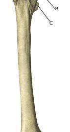
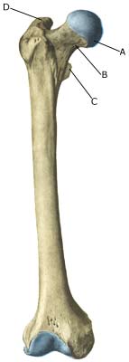
The femur is the only bone in the thigh. Innumerable muscles are attached to the bone.

  1. Caput femoris
  2. Collum femoris
  3. Trochanter minor
  4. Trochanter major

THIGH BONE FROM THE FRONT

Cause: Repeated loads, particularly when walking or running, can cause such great stress that cracks (stress fractures) appear in the shaft of the femur (article).

Symptoms: Pain and tired sensation in the thigh. The pain is aggravated upon applying pressure (direct and indirect tenderness) and applying load (walking, running).

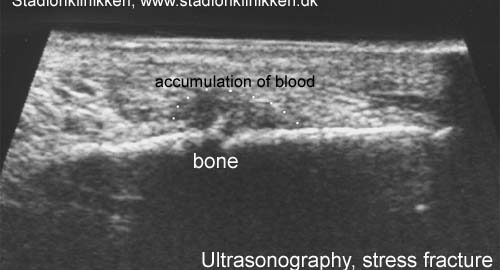
Examination: X-ray. Since many stress fractures are not visible early in the course, x-ray examination can be repeated after a few weeks, if stress fractures are still suspected. Scintigraphy, CT, MRI and ultrasound scans can often diagnose stress fractures far earlier than x-rays (Ultrasonic image). The frequency of stress fractures in the femur is probably more often than presumed (article). It is crucial for the result of the treatment that the diagnosis is made as early as possible (article).

Treatment: The treatment primarily comprises relief. Only in special cases is surgery necessary (article).

Rehabilitation: The rehabilitation is completely dependant on the type of fracture and the treatment (relief or surgical). A rehabilitation period of 2-4 months must be expected before maximum participation in sports activity can be resumed (article).

Complications: If progress is not smooth, you should be medically re-evaluated to ensure that the fracture is healing according to plan. In some cases a false joint is formed (pseudoarthrosis), which requires surgical treatment.

cause-article2

SportNetDoc

Femoral stress fractures in children.

Meaney JE, Carty H. Skeletal Radiol 1992;21(3):173-6

Stress fractures reported in the medical literature almost exclusively affect young athletes, military recruits and patients with metabolic bone disease. The classification of stress-induced bone injury is somewhat confused and includes “fatigue” stress fractures which occur in previously normal bones and “insufficiency” stress fractures which occur in bones weakened by various causes. Femoral stress fractures in children are extremely rare, and we report 5 cases in young patients who sustained their injuries during the course of normal play activities. It is well-known that these lesions can simulate malignant lesions; however, we believe that careful review of the radiographs in the context of the clinical history can in many cases lead to the correct diagnosis being made and obviate the need for further intervention.

cause-article1

SportNetDoc

Fractures of the femoral neck in children: complications and their treatment

Chladek P, Trc T. Acta Chir Orthop Traumatol Cech 2002;69(1):31-4

PURPOSE OF THE STUDY: Fractures of femoral neck in children are rare skeletal injuries which are, however, associated with a high percentage of complications. The aim of the retrospective study is to evaluate a group of patients in relation to the incidence of individual complications and demonstrate the methods used for the solution of these complications. MATERIAL: Twenty patients with fractures of femoral neck (average age 10 years 5 months) were treated at the Department of the authors in the period between 1983 and 1997. Of this 18 patients were operated on (most frequently used was internal fixation by 2 cancellous screws). In the given period 10 complications in total were handled of which 3 patients were admitted to the Department due to a complication which occurred after the surgical treatment of the fracture at another department. The group included neither pathological fractures nor patients with hormone-based slipped capital femoral epiphysis. METHODS: Fresh fractures of femoral neck were classified after Boitzy or Delbet-Collona. The results of the treatment of both fresh fractures and complications were evaluated after the interval of more than three years. RESULTS: Of 20 cases treated primarily at our Department 13 patients healed completely. Complications occurred in 10 cases (43.5% of all treated cases). The most frequent complication was avascular necrosis of femoral head (17.4% of all treated cases). This complication was managed twice by proximal femoral osteotomy, and once by arthrodesis of the hip joint and once by a triple pelvis osteotomy. The evaluation of complications shows that the more medial the neck fractured the greater was the risk of the incidence of some complication. DISCUSSION: The percentage of individual types of fractures is comparable with the results presented in the cited works, only Cheng presents an unusually share of fractures of type II to the debridement of type III. The incidence of avascular necrosis is relatively lower in the given group as compared to the cited works (Mayr states 34.6% in patients operated on). Similarly skeptical is the evaluation of fractures of type I also in other authors (Pape). CONCLUSIONS: The authors recommend as a method of choice in these fractures an early surgical treatment with emphasis on exact anatomical reduction of fragments combined with intraarticular decompression by arthrotomy in case of the presence of hemarthrosis.