DEGENERATIVE ARTHRITIS IN THE HAND
|
||
|
||
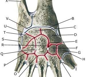
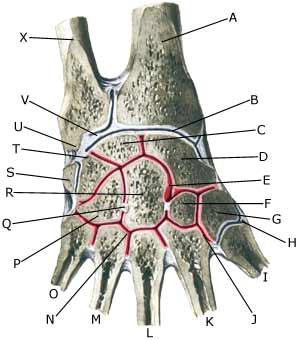
| Cause: In case of repeated loads the cartilage, primarily, and subsequently the bone beneath the cartilage, can be damaged (degenerative arthritis). The degenerative changes can in some cases cause an inflammation of the synovial membrane (synovitis), which implies fluid formation, swelling, movement constriction and pain in the joint. Degenerative changes in the hand often occur after earlier injuries (bone fractures, sprains). Degenerative changes are most frequently seen in the wrist itself (articulatio radiocarpale) or corresponding to the thumbs root joint (articulatio carpometacarpale pollicis) and in the outer joint of the finger (DIP-joint)
Symptoms: Pain in the joint upon movement. Occasional swelling in the joint (synovitis). Examination: Often an ordinary clinical examination is sufficient, although it may be necessary to supplement with an x-ray examination. Ultrasound is well suited to detect fluid in the joints (Ultrasonic image) (article). Treatment: Relief from pain inducing activities until the swelling has decreased. Rehabilitation can subsequently be commenced with the primary goal to strengthen the muscles around the joint and maintain joint-mobility. There is no treatment that can regenerate the destroyed cartilage (and bone). Cartilage transplants are not yet suitable for general degenerative changes. In cases of swelling in the joint, you can attempt to dampen the inflammation (synovitis) with rheumatic medicine (NSAID) or by draining the joint fluid and injecting corticosteroid, which can advantageously be done with ultrasound guidance. Pain with no swelling is best treated with paracetamol. Rehabilitation: The rehabilitation is dependant on which joint has suffered degenerative changes. Exercise is generally advised to maintain joint mobility and non-strenuous strength training for the muscles around the joint, which, however, does not have as large an effect as around joints with large muscles (e.g. the knee). Bandage: With degenerative changes in the wrist and thumb a bandage that supports (and relieves) the joint, can be manufactured. With degenerative changes in the thumbs base joint (MCP joint), a tape can be applied (tape-instruction) . Complications: With severe degenerative changes with pain when resting (at night) it may become necessary to fix the joint by an operation. It should be considered whether the swelling in the joint is not part of a general rheumatic disorder. |


