|
||
|
||
|
||
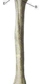
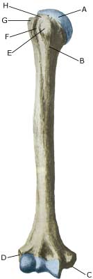
| Cause: A fracture of the arm can occur in cases of a direct fall on an outstretched arm or following a blow. In some cases the fracture occurs in the growth zone, whereas in other cases the bone “bends” without breaking (“green-stick fracture”). The most common fractures on the upper arm are fractures in the growth zone at the shoulder joint (epiphysis colli humeri), spiral fracture and green-stick fracture in the middle of the upper arm, different types of fractures around the elbow (fractura supracondylaris humeri, fractura epicondyli humeri, fractura condyli lateralis humeri, fractura capitulum humeri, fractura colli radii), fracture and green-stick fracture on the forearm and fracture in the growth zone at the wrist (epiphysis).
Symptoms: Sudden pain and pain induced constriction of movement of the arm after a fall or blow. From time to time, angular deformity of the arm can be observed. Acute treatment: Relief until a medical examination can be performed. Examination: Sudden, powerful pains in the arm with constriction of movement after a fall or blow, should always lead to acute medical examination. The fracture is usually visible on x-rays, and on the basis of the type of fracture, the correct treatment can be determined. Fractures can, however, not always be seen on x-rays, and if there is continued suspicion of a fracture, the examination should be supplemented by ultrasound scanning or MR imaging (article). The fracture can be particularly difficult to see on an x-ray if occurring in the growth zone. Treatment: Depending on the type of fracture, a choice can be made between relief and possible use of bandaging or an operation (article-1) (article-2) (article-3) Rehabilitation of children and adolescents: The type of training and rehabilitation that can be permitted is dependant upon the severity of the fracture and the type of treatment chosen. Rehabilitation should therefore take place in close cooperation with the medical staff controlling the treatment. Complications: In the vast majority of cases the fracture heals without complications, although in some cases a delayed healing occurs, possibly with the development of a false joint (pseudoartrosis), growth disturbance and possibly reduced function ability of the arm (article). In other cases, the fracture can cause sensory disturbances or affect the blood supply to the arm, which can require (acute) operation. |


