Kategoriarkiv: Arm

Nursemaid’s elbow

Diagnosis: NURSEMAID’S ELBOW
(SUBLUXATIO CAPUT RADII)


Anatomy:
The elbow joint is comprised of the upper arm bone (humerus), and one of the two forearm bones (ulna). The other forearm bone (radius) forms a joint with ulna (art. radioulnaris proximalis). A joint capsule and several strengthening ligaments surround the elbow joint.

  1. Humerus
  2. Capsula articularis
  3. Epicondylus medialis
  4. Lig. collaterale ulnare
  5. Chorda obliqua
  6. Ulna
  7. Radius
  8. Tendo m. bicipitis brachii
  9. Lig. anulare radii
  10. Lig. collaterale radiale
  11. Epicondylus lateralis

ELBOW JOINT

Cause: With a pull on the arm, the articular head of the one of the forearm bones (caput radii) can become partially dislocated. The condition is most often seen up to the age of about 5 years (article)

Symptoms: Sudden insetting pain around the elbow and pain induced restriction of mobility of the arm (article).

Examination: Sudden, strong pain in the arm with restriction of movement should always lead to a medical examination. A General Practitioner will in the vast majority of cases be able to make the diagnosis and treat the condition. X-rays or ultrasound scans are very seldom necessary (article).

Treatment: In uncomplicated cases the dislocation can normally be put into place by use of simple manipulation (article). In some circles, a couple of days of relief is subsequently recommended (article).

Rehabilitation of children and adolescents: There is not usually a need for specific rehabilitation when the dislocation has been put into place.

Inflammation of the growth zone in the elbow

Diagnosis: INFLAMMATION OF THE GROWTH ZONE IN THE ELBOW
(LITTLE LEAGUE ELBOW)


Anatomy:
The elbow joint is comprised of the upper arm bone (humerus), and one of the two forearm bones (ulna). The other forearm bone (radius) forms a joint with ulna (art. radioulnaris proximalis). A joint capsule and several strengthening ligaments surround the elbow joint. Two of the forearm’s large muscle groups are attached to the elbow, externally (lateral epicondyle), and internally (medial epicondyle), where a growth zone (apophysis) is found in adolescents which increases the risk of overload symptoms. The forearm muscles that are attached to the internal side of the elbow bend (flex) the wrist.

  1. M. biceps brachii
  2. Epikondylus mediale
  3. Aponeurosis m. bicipitis brachii
  4. M. pronator teres
  5. M. flexor carpi radialis
  6. M. palmaris longus
  7. M. flexor digitorum superficialis
  8. M. flexor carpi ulnaris

FLEXORS OF THE FOREARM

Cause: Repeated uniform loads on the forearm muscles (throwing, racket sports) cause an overload conditional inflammation with a fraying out of the bone at the point of attachment of the forearm muscles in the growth zone (apophysis) internally on the elbow (medial epicondyle) (article). The mechanism behind the development of ”inflammation of the growth zone” is identical to golf elbow for adults.

Symptoms: Slowly insetting pain on the internal side of the elbow (medial epicondyle) during and after the sports activity. With prolonged discomfort the bone fastening on the elbow will become more prominent. Boys in the 10-16 year age group are especially prone to developing the symptoms (article). The symptoms normally decline when the growth zone in the elbow closes at around age 17.

Acute treatment: Click here.

Examination: Pain when applying pressure on the inside of the elbow (medial epicondyle) which is aggravated when bending the hand against resistance, and when the forearm muscle group is stretched. A medical examination is usually sufficient to be able to make the diagnosis. If there is any doubt in the diagnosis it can occasionally be necessary to supplement with an ultrasound scan (or possibly x-ray), where a fraying out of the growth zone (apophysis) can usually be observed.

Treatment: Treatment comprises relief from the pain inducing activity (throwing, racket games) (article). If the treatment is begun very quickly, the injury can in some cases heal within a few weeks. The period of relief can be considerably longer if the pain has been present for several months. Special emphasis should be put on stretching the forearm muscles. Treatment with ice can be repeated every time the rehabilitation provokes tenderness at the muscle attachment in the elbow. With repeated pain in normal everyday life, medicinal treatment in the form of rheumatic medicine in gel form NSAID can be considered in the treatment. Injection of corticosteroid has no place in the treatment (article). The sports activity can be cautiously resumed when the pain has diminished. There will often be a recurrence of the symptoms, following which a period of relief should be started immediately. During the period of relief, it will usually be sufficient to refrain from the exercises which cause most strain on the injury (throwing, over-arm smashing), whilst many other training exercises can be performed without problems. It is only in very rare cases that an operation can become necessary (article)

Rehabilitation of children and adolescents: INSTRUCTION

Dislocation of the elbow

Diagnosis: DISCLOCATION OF THE ELBOW
(LUXATIO ARTICULI CIBITI)


Anatomy:
The elbow joint is comprised of the upper arm bone (humerus), and one of the two forearm bones (ulna). The other forearm bone (radius) forms a joint with ulna (art. Radioulnaris proximalis). A joint capsule and several strengthening ligaments surround the elbow joint.

  1. Humerus
  2. Capsula articularis
  3. Epicondylus medialis
  4. Lig. collaterale ulnare
  5. Chorda obliqua
  6. Ulna
  7. Radius
  8. Tendo m. bicipitis brachii
  9. Lig. anulare radii
  10. Lig. collaterale radiale
  11. Epicondylus lateralis

ELBOW JOINT

Cause: A dislocation of the elbow can occur following a direct fall on an outstretched arm. The dislocation can in some cases be complicated by a bone fracture, vascular damage or nerve damage.

Symptoms: Sudden insetting pain around the elbow, with pain-conditional restriction of mobility of the arm following a sudden, violent load (fall).

Acute treatment:
Click here.

Examination: Sudden, strong pain in the arm with restriction of movement following a fall should always lead to acute medical examination. Acute medical assistance should be sought due to the risk of damage to blood vessels and nerves. An X-ray examination will usually reveal the dislocation and rule out bone fracture.

Treatment: The dislocation can usually, in uncomplicated cases, be put in place without the need of surgery. Some recommend a short time where bandaging is used after the dislocation has been put into place. Surgery is often necessary in cases where complications arise from the dislocation in the form of bone fracture, vascular damage or nerve damage. Rehabilitation with exercises involving movement should be commenced as soon as possible, (article-1), (article-2).

Rehabilitation of children and adolescents: INSTRUCTION

Rehabilitation can commence shortly after the dislocation is put into place (and possible bandaging has been removed) in uncomplicated cases without bone fracture, vascular damage or nerve damage. Recommendations from your doctor must be taken into consideration in the rehabilitation program if the dislocation has involved complications and has possibly required surgery.

Complications: Tears or ruptures around the elbow will in the vast majority of cases heal without complication. Some cases will experience persistent stiffness in the elbow, looseness of the elbow, calcification in the muscles surrounding the elbow and vascular or nerve damege. Dislocation of the elbow can in some cases be complicated by ligament injuries in the wrist, (article).

Sprained finger joint

Diagnosis: SPRAINED FINGER JOINT
(DISTORSIO ARTICULI DIGITI)


Anatomy:
The five fingers on a hand consist of a total of 14 small tubular finger bones (phalanges). The finger bones are held together by various ligaments. The joint capsule around the finger joints are reinforced by three ligaments (two side ligaments and a ligament under (volar) the joint). The ligaments under the finger joints are reinforced by a small cartilage plate (fibrocartilago), that stabilizes the intermediate joint of the fingers (PIP-joints).

  1. Os metacarpale
  2. Lig. palmare
  3. Lig. collaterale
  4. Phalanx proximalis
  5. Phalanx media
  6. Phalanx distalis
  7. Capsula articularis

FINGER

  1. M. flexor digitorum superficialis
  2. Cutis (Skin)
  3. Vagina fibrosa digitorum manus
  4. Pars anularis vaginae fibrosae
  5. Pars cruciformis vaginae fibrosae

RIGHT PALM

Cause: In case of a blow, twist or over-stretching, a rupture can occur on the ligaments and cartilage discs (fibrocartilago), that surround the finger joints. In some a piece of bone can be torn off where the ligaments fasten on the side of the bone.

Symptoms: Ligament damage will typically cause a swelling of the joint and tenderness on the side of the joint which is aggravated if the joint is twisted from side to side. With damage to the cartilage disc (laesio fibrocartilaginis volaris) under the joint, the pain will be localized to the underside of the joint (volar) which is aggravated with passive over-stretching and maximal flexing of the joint.

Acute treatment: Click here.

Examination: With sudden powerful pain in a finger it is advisable to have the joint examined to ensure a correct diagnosis and treatment. The results are best if the diagnosis is made and the treatment is commenced immediately following the injury (article). A tearing of the ligament anchor on the side of the finger is occasionally visible on x-rays.

Treatment: The vast majority of ligament ruptures can be treated without surgery, while other ligament ruptures with clear laxity require surgery. It may be necessary to perform an x-ray, ultrasound scan (article 1) or an MR-scan (article 2). Damage to the cartilage disc is often treated with a splint on the finger for about 3 weeks, although not all physicians agree with this (article 3).

Rehabilitation: Fitness training in the form cycling and running along with rehabilitation according to the guidelines under rehabilitation of children and adolescents in general can usually be commenced immediately. Once the pain is completely gone you can participate in sports, although handball, volleyball, basketball and similar activities should be avoided for a few additional weeks.

Bandage: It will often stabilise the joint if the damaged finger is taped to its neighbour (tape-instruction).

Complications: In the vast majority of cases the ligaments heal without complications. In some cases, particularly after a lesion of the cartilage disc under the joints, long-term discomfort can continue for as long as a year after the injury (article). In cases of persistent pain and lack of progress you should consult your doctor again.

Fracture of the arm

Diagnosis: FRACTURE OF THE ARM
(FRACTURA HUMERI)


Anatomy:
The upper arm (humerus) consists of the head (caput), the neck (collum), the long tubular bone (corpus) and the joint in the elbow (epicondyle).
The forearm bones consist of the ulna and the radius.

  1. Caput humeri
  2. Collum chirurgicum
  3. Epicondylus medialis
  4. Epicondylus lateralis
  5. Tuberculum minus
  6. Sulcus intertubercularis
  7. Tuberculum majus
  8. Collum Anatomycum

UPPER ARM FROM THE FRONT

  1. Elbow
  2. Ulna
  3. Wrist
  4. Radius

FOREARM FROM THE FRONT

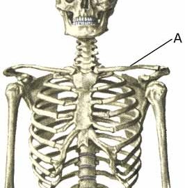
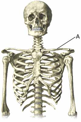
Cause: A fracture of the arm can occur in cases of a direct fall on an outstretched arm or following a blow. In some cases the fracture occurs in the growth zone, whereas in other cases the bone “bends” without breaking (“green-stick fracture”). The most common fractures on the upper arm are fractures in the growth zone at the shoulder joint (epiphysis colli humeri), spiral fracture and green-stick fracture in the middle of the upper arm, different types of fractures around the elbow (fractura supracondylaris humeri, fractura epicondyli humeri, fractura condyli lateralis humeri, fractura capitulum humeri, fractura colli radii), fracture and green-stick fracture on the forearm and fracture in the growth zone at the wrist (epiphysis).

Symptoms: Sudden pain and pain induced constriction of movement of the arm after a fall or blow. From time to time, angular deformity of the arm can be observed.

Acute treatment: Relief until a medical examination can be performed.

Examination: Sudden, powerful pains in the arm with constriction of movement after a fall or blow, should always lead to acute medical examination. The fracture is usually visible on x-rays, and on the basis of the type of fracture, the correct treatment can be determined. Fractures can, however, not always be seen on x-rays, and if there is continued suspicion of a fracture, the examination should be supplemented by ultrasound scanning or MR imaging (article). The fracture can be particularly difficult to see on an x-ray if occurring in the growth zone.

Treatment: Depending on the type of fracture, a choice can be made between relief and possible use of bandaging or an operation (article-1) (article-2) (article-3)

Rehabilitation of children and adolescents: The type of training and rehabilitation that can be permitted is dependant upon the severity of the fracture and the type of treatment chosen. Rehabilitation should therefore take place in close cooperation with the medical staff controlling the treatment.

Complications: In the vast majority of cases the fracture heals without complications, although in some cases a delayed healing occurs, possibly with the development of a false joint (pseudoartrosis), growth disturbance and possibly reduced function ability of the arm (article). In other cases, the fracture can cause sensory disturbances or affect the blood supply to the arm, which can require (acute) operation.

Fracture of the finger bones

Diagnosis: FRACTURE OF THE FINGER BONES
(FRACTURA DIGITI MANUS)


Anatomy:
The five fingers on each hand consist of a total of 14 small tubular bones (phalanges).

  1. Os lunatum
  2. Os triquetrum
  3. Os pisiforme
  4. Os hamatum
  5. Phalanx distalis
  6. Phalanx media
  7. Phalanx proximalis
  8. Os metacarpale II
  9. Ossa sesamoidea
  10. Os trapezoideum
  11. Os trapezium
  12. Os capitatum
  13. Os scaphoideum
  14. Carpus

RIGHT HAND’S BONES – PALM

Cause: With a blow, twist or fall on the hand a fracture can occur in the finger bones.

Symptoms: Sudden pain in the finger after a fall, twist or blow. Pain is aggravated upon maximal movement of the finger and upon applying direct pressure on the fracture. A visible angling of the finger can occasionally be seen.

Acute treatment: Click here.

Examination: Everyone with sudden powerful pains in a finger after a fall or blow should be examined by a doctor if the pain does not quickly abate. In case of visible angling of the finger, the medical examination should be acute. The fracture is usually visible on x-rays allowing the best treatment to be chosen accordingly.

Treatment: If there is dislocation of the finger bone, the fracture can be reset under local anaesthetic. Most fractures can be managed with relief and light bandaging, while others require a cast or surgery (article).

Rehabilitation: Fitness training in the form of cycling, running and rehabilitation according to the guidelines under rehabilitation of children and adolescents in general can usually be started immediately. When the cast is removed you can commence training of the hand and arm. Thrusts or jabs with the hand (boxing, handball, volleyball and similar sports) should be avoided for an additional few weeks.

Bandage: The fracture will often be stabilized if the finger is taped to the neighbouring finger (tape-instruction). Individual plastic bandages can be made for use during sports activity after bone fractures.

Complications: In the vast majority of cases the fracture heals without complications although in some cases the healing can complicated. Therefore, in case of persistent pain and lack of progress, you should consult your doctor again.

Fracture of the collar bone

Diagnosis: FRACTURE OF THE COLLAR BONE
(FRACTURA CLAVICULA)


Anatomy:
The collar bone (clavicula) forms a joint with the breastbone and the shoulder blade’s upper bone projection (acromion). Amongst other functions the collar bone acts as the anchor for many shoulder and chest muscles.

  1. Clavicula 

SKELETON

Cause: A fracture of the collar bone can occur in cases of a direct fall on the shoulder, or fall upon an outstretched arm; this is one of the most common fractures (5-10% of all fractures). The ligaments holding the collar bone in place may also rupture.

Symptoms: Pain in the collar bone and upon movement of the shoulder joint. In cases of a fracture with displacement of the bone, a bump is often visible on the collar bone.

Examination: Sudden, powerful pain in the shoulder with constriction of movement after a fall should always lead to acute medical examination. The fracture is usually visible on x-rays and on the basis of the type of fracture, the treatment can be determined.

Treatment: Depending on the type of fracture, rest and possibly a sling may be employed until pains decrease after a couple of weeks. In some instances, a sling is chosen to fixate the fracture (Madsens figure-8 bandage). In more complex fractures, and fractures with ruptured ligaments a surgical fixation of the fracture is most often chosen. In some cases it is an option to fixate certain fractures with a plate, which allows a faster return to sports, but the operation has a relatively high risk of complications (article).

Rehabilitation: When the pain has diminished (after 2-3 weeks) physical fitness training in the form of running may be commenced, and retraining according to the guidelines under rehabilitation of children and adolescents in general. After 4-6 weeks careful muscle training of the shoulder muscles may be started. Participation in contact sports will be possible after a couple of months have elapsed.

Complications: In the vast majority of the cases the fracture will heal without complications, although some suffer long-term discomfort, even if the fracture heals. Some fractures form a false joint (pseudoartrosis), which requires (renewed) surgery.