KNEE CAP LIGAMENT RUPTURE
|
||
|
||
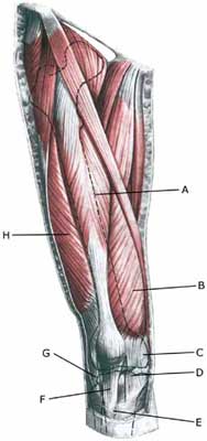
| Cause: A total or partial rupture of the knee cap ligament (ligamentum patellae) usually occurs upon an activation (contraction) of the thigh muscle simultaneously with the muscle being stretched, for example when landing, where at the same time the anterior thigh muscle is activated to set off to go quickly forward (eccentric contraction) (Photo). A basic element of total or partial ruptures of the knee cap ligament is degeneration in the ligament which has occurred due to repeated uniform loads on the knee cap ligament (jumping, kicking) causing microscopic ruptures at the ligament fastening on the lower edge of the knee cap. As the load often continues despite tenderness, which in the early stages diminishes after warm-up, a chronic inflammation gradually occurs in the ligament.
Symptoms: Sudden insetting pain in the knee cap ligament directly under the knee cap, where there is often a sensation of feeling and hearing a “snap”. Pain when activating the anterior thigh muscle (rising up from a squatting position, running and walking), pressure on the ligament and when stretching (anterior thigh muscle). A defect can often be felt in the ligament if a total rupture, and active stretching of the knee is impossible. Acute treatment: Click here. Examination: In all cases when there is a sense of a “snap”, or sudden shooting pains in the ligament with difficulty in stretching the knee, medical attention should be sought as soon as possible. Ultrasound scanning is used to advantage when making the diagnosis, as even considerable ruptures can be easily overlooked without the aid of ultrasound scanning. A swift diagnosis is of great importance for the result of the treatment (article). An ultrasound scan should be performed which will enable an appraisal of the extent of the changes in the ligament: total or partial rupture, inflammation in the tendon (tendonitis), scar tissue formation (tendinosis), calcification in the tendon, bursitis and inflammation of the tissue surrounding the tendon (peridenitis) (Ultrasonic image) (article). Treatment: Ruptures require swift operation as delay in making the diagnosis will cause a poor result. An injury period of 6-9 months must be anticipated before the sports activity can be resumed at the same level (article). Complications: A new ultrasound scan should be performed if no progress is in evidence to exclude a new (partial) rupture of the knee cap ligament and:
It will relatively often not be possible to resume the sports activity at the same level despite correct attempts at treatment and rehabilitation. Special: Since there is a risk that the injury can cause permanent disability, the injury should be reported to your insurance company. |


