Kategoriarkiv: Knee

Muscle ruptures aroud the knee

MUSCLE RUPTURES AROUND THE KNEE

Diagnosis: MUSCLE RUPTURES AROUND THE KNEE


Anatomy:
The majority of the muscle tendons running from the thigh to the shin bone pass the knee joint to the rear of the knee. The majority of “over-load” symptoms emanate therefore from the muscles in the hollow of the knee (popliteal space).


  1. M. biceps femoris (caput longum)
  2. M. semitendinosus
  3. Caput breve m. bicipitis femoris
  4. M. plantaris
  5. Tendo m. bicipitis femoris
  6. M. gastrocnemius
  7. M. sartorius
  8. M. gracilis
  9. M. semimembranosus

KNEE FROM THE REAR

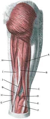
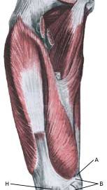
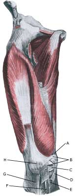
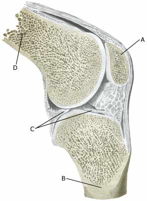
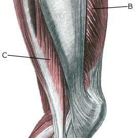
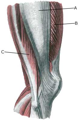
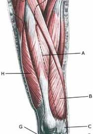
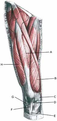
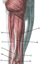
Cause: Muscle ruptures occur following sudden, violent loads, causing a total or partial rupture in the muscle or the muscle fastening. Many muscle ruptures are preceded by tenderness in the muscle or muscle tendon. Ruptures in the anterior thigh muscle fastening (tendo m bicipitis femoris) on the head of the calf bone (caput fibulae), and the calf muscle fastenings in the popliteal space are commonly seen. The vast majority of ruptures are partial ruptures.

Symptoms: Pain when applying pressure along the muscle tendon, aggravated by stretching and activation against resistance (flexing of knee). In light cases, a local tenderness can be felt after subjecting to load (“sprained muscle” “imminent pulled muscle”). In more severe cases, a sudden shooting pain can be felt in the muscle (“partial muscle rupture”, “pulled muscle”) and in the worst cases a violent “snap” can be felt, after which it is impossible to use the muscle (“total muscle rupture”). The following three symptoms are characteristic of muscle injuries: pain when applying pressure, stretching and activation against resistance (flexing of knee). A defect in the muscle can often be seen and felt in cases of a total muscle rupture, as well as swelling in the muscle (the contracted muscle belly and muscular bleeding).

Acute treatment: Click here.

Examination: A medical examination is not necessarily required in light cases. More severe cases, or cases where the treatment has not brought any improvement should be evaluated by a doctor to make a precise diagnosis. The diagnosis is usually made on the basis of a normal medical examination. Ultrasound scanning (and MR scanning) is often necessary in cases of a severe rupture in order to evaluate the extent of the rupture and bleeding (Ultrasonic image).

Treatment: Treatment comprises rest and relief, stretching and increasing loads within the pain threshold. Muscle ruptures are usually treated with a rehabilitation program, and surgery can only be considered in very rare cases with total muscle ruptures.

Complications: In case of lack of progress it should be considered whether the diagnosis is correct. Special consideration should be given to:

Luxation of the knee cap

LUXATION OF THE KNEE CAP

Diagnosis: LUXATION OF THE KNEE CAP
((Sub)luxation patellae)


Anatomy:
The large anterior thigh muscle (musculus quadriceps femoris) consists of four muscles (m vastus lateralis, m vastus medialis, m vastus intermedius & m rectus femoris). All the muscles fasten on the upper edge of the knee cap. The knee cap ligament (ligamentum patellae) connects the lower edge of the knee cap with the front part of the shin bone (tuberositas tibiae). The knee cap is held in place by the structures that fasten on the knee cap, especially the anterior thigh muscle, joint capsule and several ligaments (retiaculum patellae mediale & retiaculum patellae laterale) as well as the knee cap ligament.

  1. Tendo m. adductoris magni
  2. Retinaculum patellae mediale
  3. Meniscus medialis
  4. Ligamentum collaterale mediale/tibiale
  5. Bursa anserina
  6. Bursa subtendinea m. sartori
  7. Ligamentum patellae
  8. Patella

KNEE JOINT

Cause: Luxation of the knee cap can occur following a blow on the knee, but more often after a sudden and violent knee movement, where the knee is twisted and stretched at the same time. This can cause the knee cap to be displaced to the external side of the knee, whereby the ligaments holding the knee cap will rupture. The knee cap will often impact with the thigh bone, producing the risk of cartilage damage in the knee: cartilage damage in the joint (osteokondrale lesion), cartilage damage on the knee cap (chondromalacia patellae).

Symptoms: Sudden insetting severe pain that renders continued sports activity impossible. The knee cap can become completely displaced to the external side of the knee in some cases, and the knee will consequently be locked in a flexed position (total luxation) until the knee cap suddenly slips into place again allowing the knee to be stretched once more. In other cases, the knee cap will only be partially displaced to the external side of the knee (subluxatio patellae).

Acute treatment: Click here.

Examination: The diagnosis can be difficult if the knee cap is in its correct position, and anyone with suspicions of displacement of the knee cap should always seek medical attention to ensure the diagnosis and correct treatment. The examination will typically provoke severe pain when the knee cap is pressed outwards (lateral) whilst the knee is flexed (Apprehension test). The knee cap will often be able to be pressed further out on an injured knee than a healthy one. An MR scan will be able to reveal more information regarding the cartilage condition in the knee after a partial or total luxation of the knee cap (article).

Treatment: Partial luxation should primarily be treated with relief and rehabilitation. There is no general consensus of opinion regarding treatment of total luxation of the knee cap, some recommend surgery whilst others advocate relief and rehabilitation (article 1) (article 2). Surgery should however be considered with repeated (total or partial) luxation.

Rehabilitation of non-operated, partial luxation of the knee cap (subluxatio patellae).

Bandage: Tape and bandaging has no documented preventive effect subsequent to previous total or partial luxation of the knee cap (article), however experience has shown that this is utilised to a large degree (tape-instruction).

Complications: In case of lack of progress it should be considered whether the diagnosis is correct. Special consideration should be given to:

Special: Since there is a risk that the injury can cause permanent disability, the injury should be reported to your insurance company. A running style analysis can be considered to evaluate whether a correction to the running style can be recommended.

Degerative arthritis

DEGERATIVE ARTHRITIS

Diagnosis: DEGERATIVE ARTHRITIS
(Osteoarthritis)


Anatomy:
The bones in the knee joint comprise the thigh bone (femur), shin-bone (tibia) and the knee cap (patella). The articular surfaces of the thigh bone, shin-bone and knee cap are lined with a cartilage covering of a few millimetre’s thickness which serves to reduce the load or strain on the joint surfaces.


  1. Patella
  2. Tibiae
  3. Meniscus lateralis
  4. Femur

KNEE JOINT

Cause: Repeated and uniform loads can cause damage to the cartilage, and subsequently the bone beneath the cartilage (degenerative arthritis). of the synovial membrane (synovitis), which will cause formation of fluid, swelling, reduction in mobility and pain in the knee joint. Degenerative arthritis in the knee joint is often seen after a previous rupture of the anterior cruciate ligament, rupture of the posterior cruciate ligament eller meniscus lesion, where it has been necessary to remove the whole or part of the meniscus.

Symptoms: Pain in the joint with movement under load or strain. Difficulties in the start up phase are often experienced, alleviated after warm-up, but with pain again after a prolonged period under load. Swelling of the joint (synovitis) in occasional cases. With pronounced swelling, a fluid filled bursa can develop in the hollow (popliteal space) of the knee (Baker cyst).

Examination: Normal medical examination is usually sufficient to make the diagnosis, however, it is also often necessary to perform an x-ray (or ultrasound scan or MR scan) to make the diagnosis.

Treatment: Treatment comprises relief from the pain inducing activities until the swelling has gone down, after which training can commence with the primary aim to strengthen the muscles surrounding the joint and retain joint mobility (article). There is no treatment which can restore the damaged cartilage (and bone). Cartilage transplants are, as yet, not suitable for general degenerative arthritis. In cases of swelling in the joint (and popliteal space), inflammation of the synovial membrane (synovitis) can be attempted subdued by using rheumatic medicine (NSAID), or by draining the fluid and injecting corticosteroid. The injections can be performed to advantage by utilising an ultrasound guided method (Ultrasonic image). Pain without swelling of the joints is best treated with paracetamol. In severe cases of degenerative arthritis where there is pain when resting (at night), it may be necessary to replace the joint.

Rehabilitation: Rehabilitation is primarily aimed at strengthening the thigh muscles, whereby the joint can be stabilised and relieved to a certain extent.

Complications: Degenerative arthritis which is positioned on the weight-bearing parts of the joint surfaces represents one of the most serious sports injuries, and often results in an end to the sporting career. It is usually possible to participate in sports with lesser knee straining activity (i.e. swimming), whereas sports with great knee straining activity (i.e. running, football) should be treated with reservation.

Special: Shock absorbing shoes or inlays will reduce the load on the knee.

Cartilage damage in the joint

CARTILAGE DAMAGE IN THE JOINT

Diagnosis: CARTILAGE DAMAGE IN THE JOINT
(Osteochondral lesion)


Anatomy:
The bones in the knee joint comprise the thigh bone (femur), shin-bone (tibia) and the knee cap (patella). The articular surfaces of the thigh bone, shin-bone and knee cap are lined with a cartilage covering of a few millimetre’s thickness which serves to reduce the load or strain on the joint surfaces.

Cause: Twists in the knee joint causing the thigh bone and shin bone to collide can cause damage to the cartilage in the knee. In some cases a piece of cartilage can be shed which can wander in the joint (joint-mouse) and become inflamed and increase the production of synovial fluid.

Symptoms: Pain in the joint when under load or strain. Often, periodic swelling of the joint (synovitis).

Examination: Normal medical examination is often not sufficient. To make the diagnosis correctly it is therefore necessary to perform an arthroscopic examination (telescopic examination of the joint (Photo) or an MR-scanning  (Ultrasonic image).

Treatment: Treatment comprises relief from the painful activities until the pain is no longer experienced, after which gradual re-training can be commenced. There is no treatment that can restore the damaged cartilage, which has itself poor restorative ability. Different procedures to enhance the healing can be attempted using arthroscopy, however, the results are generally unsatisfactory. Results from experimental cartilage transplants are still not successful enough to warrant introduction as a routine treatment in the near future (article). Joint-mouse that provokes the symptoms must be surgically removed. Injection of corticosteroid in the knee joint, performed under ultrasound guidance, can be attempted to combat prolonged and pronounced cases of fluid accumulation in the knee joint (synovitis).

Rehabilitation: Rehabilitation is completely dependent upon the type of cartilage damage (size and position in the joint) and treatment (conservative or surgical). It is generally attempted to strengthen the musculature around the knee joint.
Also read rehabilitation, general.

Complications: Large cartilage damage which is positioned on the weight-bearing parts of the joint represents one of the most serious sports injuries, and often results in an end to the sporting career.

Special: As there is a risk that the injury can cause permanent disability, all cases should be reported to your insurance company.

Cartilage damage on the knee cap

BRUSKBESKADIGELSE BAGPÅ KNÆSKALLEN

Diagnosis: CARTILAGE DAMAGE ON THE KNEE CAP
(Chondromalacia patellae)


Anatomy:
The bones in the knee joint comprise the thigh bone (femur), shin-bone (tibia) and the knee cap (patella). The articular surfaces of the thigh bone, shin-bone and knee cap are lined with a cartilage covering of a few millimetre’s thickness which serves to reduce the load or strain on the joint surfaces.

  1. Patella
  2. Tibiae
  3. Meniscus lateralis
  4. Femur

KNEE JOINT

Cause: Repeated and uniform loads can cause damage to the cartilage, and subsequently the bone beneath the cartilage (degenerative arthritis). Degenerative arthritis changes (osteoarthrosis) on the rear of the knee cap occur often after a fall on the knee and with many smaller over-loads, however, in many cases the cause is unknown. A weak thigh muscle and increased outward turning of the foot (pronation) have been suspected of being a contributory factor to the disease.

Symptoms: Pain in the joint when under load or strain, especially with bent knee (for example stairway steps). A sensation of stiffness in the knee after a prolonged period of sitting. Swelling of the joint (synovitis) in occasional cases.

Examination: Normal medical examination is often sufficient. A characteristic of the condition is that the pain occurs when the knee cap presses against the thigh bone, producing a rough, grating sensation. If there are any doubts regarding the diagnosis the examination can be supplemented by X-rays, arthroscopic examination or an MR-scan.

Treatment: Treatment comprises relief from the painful activities until the pain is no longer experienced, after which gradual re-training can be commenced primarily aimed at strengthening the thigh muscles. There is no treatment that can restore the damaged cartilage on the knee cap. Several different surgical treatments have been attempted with unsatisfactory results (article).

Bandage: Some patients have felt that the discomfort from cartilage damage behind the knee cap has been helped by applying a knee bandage that holds the knee cap slightly to the side. Alternatively, knee cap stabilising tape can be used (tape-instruction).

Complications: If smooth progress is not achieved it should be considered whether the diagnosis is correct, which will often require supplementary examination (X-ray, ultrasound scanning, MR scanning or arthroscopy). The following should especially be considered:

It is usually possible to participate in sports with lesser knee straining activity (i.e. swimming) without great discomfort despite a large degree of cartilage damage, whereas sports with great knee straining activity (i.e. running, football) should be treated with reservation.

Specielt: Shock absorbing shoes or inlays will reduce the load in the knee. In case of lack of progress or recurrence after successful rehabilitation, running style analysis can be considered to evaluate whether correction of the running style can be recommended.

Bone membrane tear

BONE MEMBRANE TEAR

Diagnosis: BONE MEMBRANE TEAR
(Perisotael avulsion)


Anatomy:
The bones in the knee joint comprise the thigh bone (femur), shin-bone (tibia) and the knee cap (patella). A small joint is also formed between the shin-bone and the calf bone (fibula).


  1. Patella
  2. Tibiae
  3. Meniscus lateralis
  4. Femur

KNEE JOINT

Cause: A violent twist in the knee joint can stretch the ligaments and tear a small piece of bone membrane from the ligament attachment point. In the majority of cases the symptoms will diminish after a few weeks, however, in some cases the tears will provoke an inflammation and can be of a longer duration. Bone membrane tears in the knee joint area are common, and can be seen in most cases where the person has played football for many years (“football-knee”). Bone membrane tears can occur on all the bones of the knee where tendons or ligaments are attached, but is most commonly seen on the inner and outer part of the knee joint as a consequence of previous spraining of the collateral ligaments.

Symptoms: Pain when applying pressure, and when stretching the tendon or ligaments which are attached to the bone where the tear has occurred.

Acute treatment: Click here.

Examination: Normal clinical examination is often sufficient. Larger tears can be seen on an X-ray. Many lesser tears can be best seen via an ultrasound scan, from which an inflammation surrounding the tear can also be seen (Ultrasonic image).

Treatment: Minor tears merely require relief from the pain inducing activities. Larger tears can require surgery. Some cases can cause prolonged discomfort with pain that does not recede despite relief. This can be due to the tear causing chronic inflammation in the tissue. In such cases, rheumatic medicine (NSAID) or injection of corticosteroid in the area surrounding the tear can be recommended.

Rehabilitation: Rehabilitation is totally dependent upon the type of tear, and the treatment (conservative or surgical). The tears on the inner side of the knee are usually re-trained in the manner of inner collateral ligaments ruptures, whilst tears externally to the knee are re-trained asouter collateral ligament ruptures.
Also read rehabilitation, general.

Complications: If smooth progress is not achieved it should be considered whether the diagnosis is correct, which will often require supplementary examination (X-ray, ultrasound scanning or MR scanning). The following should especially be considered:

Bone fracture in the knee

KNOGLEBRUD I KNÆET

Diagnosis: BONE FRACTURE IN THE KNEE


Anatomy:
The bones in the knee joint comprise the thigh bone (femur), shin-bone (tibia) and the knee cap (patella). A small joint is also formed between the shin-bone and the calf bone (fibula).

  1. Patella
  2. Tibiae
  3. Meniscus lateralis
  4. Femur

KNEE JOINT

Cause: A bone fracture is usually caused by a blow or violent twist.

Symptoms: Sudden insetting pain which is aggravated when applying pressure and when applying load or strain. The symptoms are dependant upon which bone is broken. In cases where the knee cap is cracked right across, the stretching function will discontinue.

Acute treatment: Click here.

Examination: Medical assistance (casualty ward) should be sought immediately if there are any suspicions of a bone fracture in order for the diagnosis to be confirmed by X-ray examination. A fracture in the growth zone surrounding the knee can be difficult to determine in children and adolescents.

Treatment: Treatment is completely dependent upon the type of fracture, and can consist of relief, bandage/plaster cast or operation.

Rehabilitation: Rehabilitation is totally dependent upon the type of fracture, and the treatment (relief, bandage/plaster cast or operation). All attempts must be made to preserve the muscle strength in the thigh and shin bone as far as possible, and the mobility of the knee joint.
Also read rehabilitation, general.

Complications: If there is not a steady improvement in the condition a medical examination should be performed once more to ensure that the fracture is healing according to plan..

Specielt: As there is a risk that the injury can cause permanent disability, all cases should be reported to your insurance company.

Runner’s knee

RUNNERS KNEE

Diagnosis: RUNNER’S KNEE


Anatomy:
A strong tendon (tractus iliotibialis) runs externally from the hip crest to the shin-bone, upon which many the thigh muscles fasten.


  1. Tractus iliotibialis
  2. M. vastus lateralis
  3. M. biceps femoris

KNEE, OUTER THIGH

Cause: Repeated uniform movement in the knee joint (running, cycling) can cause the tendon (iliotibial tract) to slide over the external side of the thigh bone directly above the knee. An inflammation can consequently occur in the tendon, or in the bursa directly underneath. Athletes with a tendency to bowlegs have an increased risk (Photo)

Symptoms: Slowly insetting pain externally on the knee, aggravated when applying pressure, when stretching the iliotibial tract and running.

Acute treatment: Click here.

Examination: The diagnosis can usually be made on the basis of a normal medical examination. If there are any doubts regarding the diagnosis, an ultrasound scan (or MR scan) is recommended.

Treatment: Treatment comprises relief, stretching of the iliotibial tract and rehabilitation. It is imperative that footwear has good shock absorbing soles. An incorrect foot posture should be corrected by use of special shoes or insoles. If progress is not forthcoming, medical treatment in the form of rheumatic medicine (NSAID) or the injection of corticosteroid can be considered. A surgical splitting of the ligament can be performed in sever cases which do not react to relief, correct rehabilitation or medicinal treatment (article).

Complications: If smooth progress is not achieved it should be considered whether the diagnosis is correct, which will often require supplementary examination (X-ray, ultrasound scanning or MR scanning). The following should especially be considered:

Special: Shock absorbing shoes or inlays will reduce the load in the knee. In case of lack of progress or recurrence after successful rehabilitation, a running style analysis can be considered to evaluate whether correction of the running style is indicated.

Osgood-Schlatter disease

OSGOOD-SCHLATTER DISEASE

Diagnosis: OSGOOD-SCHLATTER DISEASE


Anatomy:
The large anterior thigh muscle (musculus quadriceps femoris) consists of four muscles (m vastus lateralis, m vastus medialis, m vastus intermedius & m rectus femoris). All the muscles fasten on the upper edge of the knee cap. The knee cap ligament (ligamentum patellae) connects the lower edge of the knee cap with the front part of the shin bone (tuberositas tibiae), where children and adolescents have a growth zone. The function of the knee cap ligament is therefore, when the knee is stretched, to transfer the power the large thigh muscle produces.


  1. M. rectus femoris
  2. M. vastus medialis
  3. Retinaculum patellae mediale
  4. Retinaculum patellae mediale
  5. Tuberositas tibiae
  6. Lig. Patellae
  7. Retinaculum patellae laterale
  8. M. vastus lateralis

KNEE FROM THE FRONT


(Photo)

Cause: Repeated uniform loads on the knee cap (jumping, kicking) cause an over-load conditioned inflammation with a fraying of the bone at the knee cap ligaments fastening on the growth zone at the upper front part of the shin bone (tuberositas tibiae). The mechanism behind development of Osgood-Schlatter disease is the same in adults as for jumper’s knee. Osgood-Schlatter disease is one of the most common sports injuries in children and adolescents.

Symptoms: Slowly insetting tenderness at the upper, front part of the shin bone (tuberositas tibiae) during and after the sports activity. If the discomfort has a long duration, the bone fastening on the shin bone will become more prominent and can become so large that kneeling will be a problem. It is especially boys in the 10-16 age group that have the symptoms, and the condition is very common and can be seen in almost all boys’ football teams. The symptoms will diminish at around age 17 when the growth zone on the shinbone closes.

Acute treatment: Click here.

Examination: The diagnosis can usually be made with certainty under a normal medical examination, revealing localised tenderness on the knee cap fastening on the upper, front part of the shin bone. If there is any doubt surrounding the diagnosis an ultrasound scan can be performed to identify the changes (Ultrasonic image), however, this is seldom necessary in uncomplicated cases (article).

Treatment: The treatment comprises relief from the pain inducing activity (jumping, kicking). The injury can in some cases heal within a few weeks if the treatment is instigated quickly whereas a rehabilitation period of several months must be anticipated if the pain has been in evidence for some months. Emphasis is placed on stretching of the anterior thigh muscle. Ice treatment can be repeated every time tenderness is provoked in the knee cap ligament fastening during the rehabilitation period. If during the rehabilitation period pain is experienced when walking, medicinal treatment in the form of rheumatic medicine (NSAID) in gel form. Injection of corticosteroid should not be considered in the treatment (article). The sports activity can be cautiously resumed when the pain has diminished. Relapses will often occur, which should be followed as soon as possible with a period of relief. During the relief period it will usually be sufficient to abstain from the most stressful exercises (jumping), whilst many other training exercises can be performed without discomfort. In the majority of cases it will be therefore be possible to participate in at least a part of the sports activity (for example as goal-keeper).

Bandage: Some patients have the opinion that the application of tape or bandage around the shin bone directly under the knee cap can relieve the discomfort (tape-instruction).

Complications: In case of lack of progress it should be considered whether the diagnosis is correct or whether complications have arisen. Special consideration should be given to:

Kun i meget sjældne tilfælde er der beskrevet afrivning af knæskalssenen fra fæste på skinnebenet. Den afrevne knogledel (hvorpå knæskalssenen fæster) kan ved en operation skrues fast på skinnebenet igen. (article).

Special: Shock absorbing shoes or inlays will reduce the load on the knee.

Knee cap ligament rupture

KNEE CAP LIGAMENT RUPTURE

Diagnosis: KNEE CAP LIGAMENT RUPTURE
(Ruptura ligamentum patellae)


Anatomy:
The large anterior thigh muscle (musculus quadriceps femoris) consists of four muscles (m vastus lateralis, m vastus medialis, m vastus intermedius & m rectus femoris). All the muscles fasten on the upper edge of the knee cap. The knee cap ligament (ligamentum patellae) connects the lower edge of the knee cap with the front part of the shin bone (tuberositas tibiae). The function of the knee cap ligament is therefore, when the knee is stretched, to transfer the power the large thigh muscle produces.


  1. M. rectus femoris
  2. M. vastus medialis
  3. Retinaculum patellae mediale
  4. Retinaculum patellae mediale
  5. Tuberositas tibiae
  6. Lig. Patellae
  7. Retinaculum patellae laterale
  8. M. vastus lateralis

KNEE FROM THE FRONT

Cause: A total or partial rupture of the knee cap ligament (ligamentum patellae) usually occurs upon an activation (contraction) of the thigh muscle simultaneously with the muscle being stretched, for example when landing, where at the same time the anterior thigh muscle is activated to set off to go quickly forward (eccentric contraction) (Photo). A basic element of total or partial ruptures of the knee cap ligament is degeneration in the ligament which has occurred due to repeated uniform loads on the knee cap ligament (jumping, kicking) causing microscopic ruptures at the ligament fastening on the lower edge of the knee cap. As the load often continues despite tenderness, which in the early stages diminishes after warm-up, a chronic inflammation gradually occurs in the ligament.

Symptoms: Sudden insetting pain in the knee cap ligament directly under the knee cap, where there is often a sensation of feeling and hearing a “snap”. Pain when activating the anterior thigh muscle (rising up from a squatting position, running and walking), pressure on the ligament and when stretching (anterior thigh muscle). A defect can often be felt in the ligament if a total rupture, and active stretching of the knee is impossible.

Acute treatment: Click here.

Examination: In all cases when there is a sense of a “snap”, or sudden shooting pains in the ligament with difficulty in stretching the knee, medical attention should be sought as soon as possible. Ultrasound scanning is used to advantage when making the diagnosis, as even considerable ruptures can be easily overlooked without the aid of ultrasound scanning. A swift diagnosis is of great importance for the result of the treatment (article). An ultrasound scan should be performed which will enable an appraisal of the extent of the changes in the ligament: total or partial rupture, inflammation in the tendon (tendonitis), scar tissue formation (tendinosis), calcification in the tendon, bursitis and inflammation of the tissue surrounding the tendon (peridenitis) (Ultrasonic image) (article).

Treatment: Ruptures require swift operation as delay in making the diagnosis will cause a poor result. An injury period of 6-9 months must be anticipated before the sports activity can be resumed at the same level (article).

Complications: A new ultrasound scan should be performed if no progress is in evidence to exclude a new (partial) rupture of the knee cap ligament and:

It will relatively often not be possible to resume the sports activity at the same level despite correct attempts at treatment and rehabilitation.

Special: Since there is a risk that the injury can cause permanent disability, the injury should be reported to your insurance company.