BONE MEMBRANE TEAR
|
||
|
||
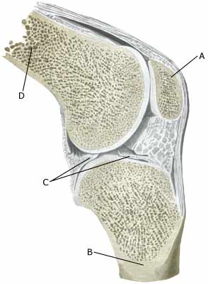
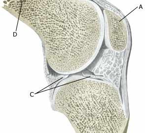
| Cause: A violent twist in the knee joint can stretch the ligaments and tear a small piece of bone membrane from the ligament attachment point. In the majority of cases the symptoms will diminish after a few weeks, however, in some cases the tears will provoke an inflammation and can be of a longer duration. Bone membrane tears in the knee joint area are common, and can be seen in most cases where the person has played football for many years (“football-knee”). Bone membrane tears can occur on all the bones of the knee where tendons or ligaments are attached, but is most commonly seen on the inner and outer part of the knee joint as a consequence of previous spraining of the collateral ligaments.
Symptoms: Pain when applying pressure, and when stretching the tendon or ligaments which are attached to the bone where the tear has occurred. Acute treatment: Click here. Examination: Normal clinical examination is often sufficient. Larger tears can be seen on an X-ray. Many lesser tears can be best seen via an ultrasound scan, from which an inflammation surrounding the tear can also be seen (Ultrasonic image). Treatment: Minor tears merely require relief from the pain inducing activities. Larger tears can require surgery. Some cases can cause prolonged discomfort with pain that does not recede despite relief. This can be due to the tear causing chronic inflammation in the tissue. In such cases, rheumatic medicine (NSAID) or injection of corticosteroid in the area surrounding the tear can be recommended. Rehabilitation: Rehabilitation is totally dependent upon the type of tear, and the treatment (conservative or surgical). The tears on the inner side of the knee are usually re-trained in the manner of inner collateral ligaments ruptures, whilst tears externally to the knee are re-trained asouter collateral ligament ruptures. Complications: If smooth progress is not achieved it should be considered whether the diagnosis is correct, which will often require supplementary examination (X-ray, ultrasound scanning or MR scanning). The following should especially be considered: |