|
||
|
||
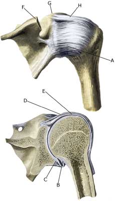
| Cause: In case of violent strain the head of the humerus (caput) may be displaced, causing a strain of the joint-capsule and the ligaments.
Symptoms: Pain corresponding to the front of the shoulder. Normal passive mobility, but often restricted active mobility. Acute treatment: Click here. Examination: Light cases require no immediate treatment, but in case of powerful pain and movement constriction, or in lack of progress, a medical examination should be performed for a precise diagnosis. It may be necessary to supplement with further examinations (X-ray, MRI (article), or ultrasound). In some cases the injury is combined with a meniscus lesion in the shoulder (laesio labrum glenoidale). Treatment: Most cases heal in a few weeks with relief and carefully increasing load within the pain threshold. Rehabilitation is aimed at strengthening the muscles around the shoulder joint (article). Complications: If satisfactory progress is not made, a physician should be consulted to ensure that the diagnosis is correct and that no complications have arisen. Amongst others the following should be considered: |


