Kategoriarkiv: Shoulder

Nerve entrapment on the back of the shoulder blade

NERVEAFKLEMNING BAG PÅ SKULDERBLADET

Diagnosis: NERVE ENTRAPMENT ON THE BACK OF THE SHOULDER BLADE
(Entrapment nervus suprascapularis)


Anatomy:
Behind the upper edge of the shoulder blade in a small channel (incisura scapulae) closed by a ligament (ligamentus transversum), runs a nerve (N suprascapularis) that distributes nerve fibres to parts of the shoulder joint-capsule and the upper and lower shoulder blade muscle (M supraspinatus and M infraspinatus)

  1. Acromion
  2. Cavitas glenoidalis
  3. Fossa supraspinata
  4. Spina scapulae
  5. Incisura scapulae
  6. Processus coracoideus

RIGHT SHOULDER BLADE SEEN
FROM THE REAR

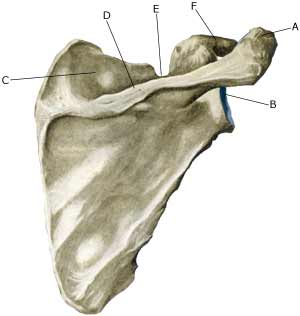
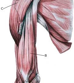
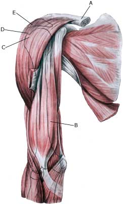
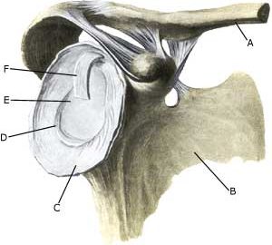
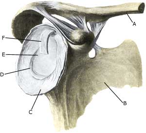
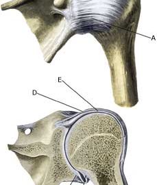
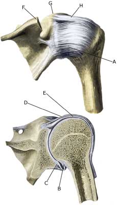
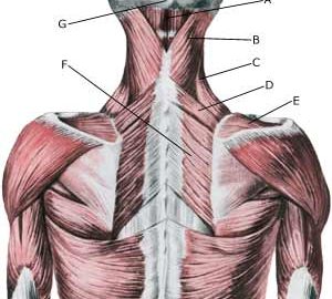
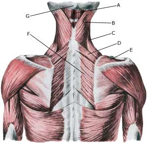
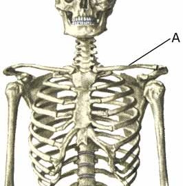
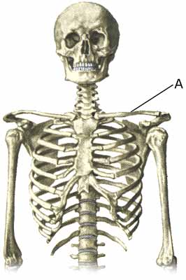
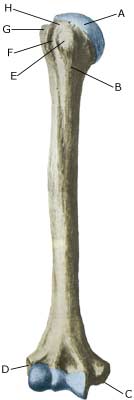
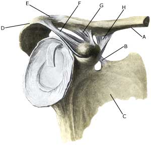
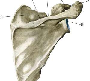
Cause: In case of repeated strain with movement in the shoulder joint, an entrapment of the nerve (article). Entrapment of the nerve may also occur as the result of a blow.

Symptoms: Usually a burning or cutting pain in the shoulder. Possibly a sense of weakness in the shoulder. In case of long-term symptoms degradation of the shoulder blade muscles will occur.

Examination: Uncharacteristic shoulder symptoms, not improved by relief, should be examined by a physician. If the results of the examination indicate nerve entrapment the diagnosis is made by a nerve conduction velocity test (EMG).

Treatment: The treatment involves relief from the pain inducing activity, stretching and rehabilitation of the muscles around the shoulder. If no progress is made with relief and rehabilitation, surgical intervention may be attempted (article-1) (article-2).

Relief is the primary element, and it is particularly important that the rehabilitation does not cause any discomfort. Fitness training should not be resumed until the pain has stopped

Complications: If satisfactory progress is not made, a physician should be consulted to ensure that the diagnosis is correct and that no complications have arisen. Amongst others the following should be considered:

Impigment syndrome in the shoulder

IMPINGEMENT SYNDROME IN THE SHOULDER

Diagnosis: IMPINGEMENT SYNDROME IN THE SHOULDER
(Impingement)


Anatomy:
Four muscles are part of the rotator cuff surrounding the shoulder joint, and controls, coordinates and assists movement of the shoulder: M supraspinatus (the upper shoulder blade muscle), M infraspinatus (the lower shoulder blade muscle), M subscapularis and M teres minor. When the arm is moved away from the body and above the head (abducated) the supraspinatus muscle slips under the upper bone projection of the shoulder blade (acromion).


  1. Clavicula
  2. M. biceps brachii
  3. Tuberculum majus
  4. M. deltoideus
  5. Acromion

(Photo)

SHOULDER AND UPPER ARM MUSCLES
FROM THE FRONT

Cause: There are two main causes for impingement: One external and one internal.
The external impingement is usually caused by repeated loads with the arm above the head (tennis, swimming) and causes an inflammation in the upper shoulder blade muscle (M supraspinatus) and the above-lying bursa (bursae subacromialis), which swells and may become squeezed between the head of the humerus (caput humeri) and the upper bone projection of the shoulder blade (acromion), when the arm is raised above the head (see inflammation of the bursa).
The internal impingement is caused by the supraspinatus muscle tendon scraping against the edge of the shoulder’s meniscus (posterior-superior edge of the labrum glenoidale) when throwing (90 degrees abduction and maximal extension and outwards rotation), causing damage to both the muscle tendon and the meniscus (article).

Symptoms: Slowly insetting local tenderness after the load, externally and to the front of the shoulder. Sometimes radiating down the upper arm. Pain deteriorates when external pressure is applied to the supraspinatus muscle, on the front of the shoulder and when the muscle is brought into contact with the meniscus in the shoulder (throwing exercises) or when the arm is brought approx. 80-120 degrees away from the body (abduction).

Acute treatment: Click here.

Examination: If no progress is made with relief and discontinuance of the pain inducing activity, a medical examination should be carried out to ensure that the diagnosis is correct and to commence the correct treatment. It is important to make the diagnosis as fast as possible, since the injury may otherwise slowly deteriorated (article). Ordinary medical examination is usually sufficient to make the diagnosis (internal impingement-test: Chicken-wing test), but if there is uncertainty concerning the diagnosis, it should be supplemented with an ultrasound scan, which is the most suitable examination for shoulder injuries (article). Though it is not rare for the diagnosis only being made after a arthroscopic examination is carried out (telescopic examination of the shoulder joint).

Treatment: The treatment primarily involves relief from the pain inducing activity, stretching and rehabilitation of the muscles around the shoulder, Impingement syndrome in athletes. In case of lack of progress a medical treatment can be considered, rheumatic medicine (NSAID) or injection of corticosteroid (usually in the subacromiale bursa and possibly in the shoulder joint itself as well (internal impingement with signs of synovitis)). Since the injection of corticosteroid is part of a long-term rehabilitation of a long-term injury, it is often necessary for the rehabilitation period to stretch over several weeks to months in order to reduce the risk of recurrences. Ultrasound guided injections offer the optimal effect with a minimum of risk, (article). In case of lack of progress in the rehabilitation and medical treatment, an operative treatment can be attempted.

Complications: If progress is not smooth, it should be considered whether the diagnosis is correct or whether complications have arisen. Amongst others the following should be considered:

Meniscus lesion in the shoulder

MENISCUS LESION IN THE SHOULDER

Diagnosis: MENISCUS LESION IN THE SHOULDER
(Laesio labrum gleniodale)


Anatomy:
Along the edge of the articular surface (cavitas glenoidalis) in the shoulder is a meniscus (labrum glenoidale). The meniscus is important to the stability of the shoulder.


  1. Clavicula
  2. Scapula
  3. Capsula articularis
  4. Labrum glenoidale
  5. Cavitas glenoidalis
  6. M. biceps brachii
    (caput longum)

ARTICULAR SURFACE OF THE SHOULDER
BLADE FROM THE FRONT

Cause: In case of repeated (over) loading or in case of one excessive load, the meniscus (labrum glenoidale) may rupture or be ripped from its attachment on the articular surface. After dislodging of the meniscus it may be squeezed in the joint. Lesion of the meniscus is often combined with (partial) ruptures of the shoulder blade muscles: Rupture of the upper shoulder blade muscle, Rupture of the lower shoulder blade muscle and in case of luxation in the shoulder and may occur as the result of a fall, where you break the fall with an outstretched arm or if handball players are “grabbed in the arm” in the moment of shooting (article).
There are several different meniscus lesions.

Symptoms: Pain in the shoulder joint with certain movements. A sense of something “slipping” in the joint is common.

Acute treatment: Click here.

Examination: A suspicion of a meniscus lesion in the shoulder (“slipping sensation”) and lack of progress should always lead to a medical examination (Apprehesion test, relocation test). An impression of the meniscus can be formed with MR and ultrasound scan, but the lesion is best assessed using arthroscopy (telescopic examination of the shoulder joint), where treatment can be performed at the same time.

Treatment: A referral to arthroscopy is advised where there is a suspicion of a meniscus lesion and smooth progress is not achieved with relief and rehabilitation, where it will be possible to determine the diagnosis, and whether it is possible to stitch the meniscus in place or whether parts of it should be removed. The results of the operation in cases of isolated meniscus lesion are usually good (article).

Complications: If progress is not smooth, it should be considered whether the diagnosis is correct or whether complications have arisen. Amongst others the following should be considered:

Sprained shoulder

SPRAINED SHOULDER

Diagnosis: SPRAINED SHOULDER
(Distorsio art. humeroscapularis)


Anatomy:
The shoulder joint consists of the head of the humerus (caput humeri) and the articular surface on the shoulder blade (cavitas glenoidalis). Along the edge of the articular surface on the shoulder blade is a meniscus (labrum glenoidale). The joint is surrounded by a joint-capsule, which is strengthened by numerous ligaments. The shoulder muscles surrounding the joint-capsule further strengthen the joint.

  1. Capsula articularis
  2. Capsula articularis
  3. Labrum glenoidale
  4. Capsula articularis
  5. M. biceps brachii 
    (caput longum)
  6. Acromion
  7. Processus coracoideus
  8. Ligamentum coracohumerale

SHOULDER JOINT

Cause: In case of violent strain the head of the humerus (caput) may be displaced, causing a strain of the joint-capsule and the ligaments.

Symptoms: Pain corresponding to the front of the shoulder. Normal passive mobility, but often restricted active mobility.

Acute treatment: Click here.

Examination: Light cases require no immediate treatment, but in case of powerful pain and movement constriction, or in lack of progress, a medical examination should be performed for a precise diagnosis. It may be necessary to supplement with further examinations (X-ray, MRI (article), or ultrasound). In some cases the injury is combined with a meniscus lesion in the shoulder (laesio labrum glenoidale).

Treatment: Most cases heal in a few weeks with relief and carefully increasing load within the pain threshold. Rehabilitation is aimed at strengthening the muscles around the shoulder joint (article).

Complications: If satisfactory progress is not made, a physician should be consulted to ensure that the diagnosis is correct and that no complications have arisen. Amongst others the following should be considered:

Muscle infiltrations in the neck/shoulder

MUSCLE INFILTRATIONS IN THE NECK-SHOULDER

Diagnosis: MUSCLE INFILTRATIONS IN 
THE NECK-SHOULDER


Anatomy:
Numerous muscles origin and fasten around the shoulder/neck.


  1. M. semispinalis capitis
  2. Mm. splenii capitis et cervicis
  3. M. levator scapulae
  4. M. rhomboideus minor
  5. M. supraspinatus
  6. M. rhomboideus major
  7. Protuberantia occipitalis externa

DEEP NAPE AND BACK MUSCLES FROM THE REAR

Cause: All muscles around the shoulder can become tense and tender, and develop tender or sore muscle knots (muscle infiltration). The cause is not known for certain, but it is known that incorrect working position can trigger muscle infiltration, and that can arise secondary to pain conditions other places in the neck-shoulder-girdle. The most frequent place for muscle infiltrations is the shoulder blade muscles (M supraspinatus and M infraspinatus, from which the pain can radiate down into the arm) and the large neck-back muscles (M Trapezius, M levator scapulae, M rhomboideus).

Symptoms: Tenderness in the muscles exaggerated when applying pressure on the muscle. There are often radiations to the arm (from muscle infiltrations around the shoulder blade) and to the head (tension head-ache with pressure behind the eyes) in cases of muscle infiltrations on the muscle fastenings in the back of the head.

Examination: Muscle infiltrations do not usually require closer examination, but in case of long-term discomfort the triggering cause should be ruled out.

Treatment: If possible the provoking cause must be removed or treated. The treatment of muscle infiltrations is furthermore stretching and increasing fitness training (article-1) (article-2) (article-3), (article-4).

Complications: If progress is not smooth, it should be considered whether the diagnosis is correct or whether complications have arisen. Amongst others the following should be considered:

Frozen shoulder

FROZEN SHOULDER

Diagnosis: FROZEN SHOULDER
(Periartrosis humeroscapularis)


Anatomy:
The shoulder joint consists of the head of the humerus (caput humeri) and the articular surface on the shoulder blade (cavitas glenoidalis). Along the edge of the articular surface on the shoulder blade, is a meniscus (labrum glenoidale). The joint is surrounded by a joint-capsule, which is strengthened by numerous ligaments. The shoulder muscles surrounding the joint-capsule further strengthen the joint.

  1. Capsula articularis
  2. Capsula articularis
  3. Labrum glenoidale
  4. Capsula articularis
  5. M. biceps brachii 
    (caput longum)
  6. Acromion
  7. Processus coracoideus
  8. Ligamentum coracohumerale

SHOULDER JOINT

Cause: In some cases a powerful strain (fall) can, for unknown reasons, trigger a shrinking of the joint-capsule, reducing the mobility of the joint in all directions. In other cases, the condition arises without preceding damage.

Symptoms: Usually pain in the shoulder (in many cases from an inflammation of the bursa (bursitis subacromialis)). After a few months, movement constriction of the shoulder joint occurs resulting of the reduction of all movement. After a few years (on average 2½ years) pain diminishes and after another few months mobility in the shoulder joint is normalized (article).

Examination: Anyone suspecting a frozen shoulder should be medical examined. A normal medical examination is usually sufficient in order to make the diagnosis There is typically movement constriction in all directions, including when the arm is passively rotated outwards. If the shoulder blade is held fixed, only very modest mobility will be possible.

Treatment: Since the condition is benign and usually heals spontaneously, treatments with the risk of side effects, should be used with restraint. Usually mild painkillers are recommended (paracetamol) or, if needed, rheumatic medicine (NSAID). In the absence of any effect corticosteroid can be injected into the shoulder joint and the bursa, if inflammed (article). Some have used corticosteroid in pill form. Various operations have been attempted, where the joint capsule is cut through (or split), however, further complications are often associated with these operations, so a certain amount of restraint is advised (article).

Complications: The diagnosis should be verified through a medical examination. Amongst other things consideration must be given to:

The physician will also consider an X-ray of the lungs and a glucose measurement, since the condition is described slightly more often in people with lung cancer and diabetes.

Fracture of the collar bone

FRACTURE OF THE COLLAR BONE

Diagnosis: FRACTURE OF THE COLLAR BONE
(Fractura clavicula)


Anatomy:
The collar bone (clavicula) forms a joint with the breastbone and the shoulder blade’s upper bone projection (acromion). Amongst other functions the collar bone acts as the anchor for many shoulder and chest muscles.

  1. Clavicula

SKELETON

Cause: A fracture of the collar bone can occur in cases of a direct fall on the shoulder, or fall upon an outstretched arm; this is one of the most common fractures (5-10% of all fractures). The ligaments holding the collar bone in place may also rupture.

Symptoms: Pain in the collar bone and upon movement of the shoulder joint. In cases of a fracture with displacement of the bone, a bump is often visible on the collar bone.

Examination: Sudden, powerful pain in the shoulder with constriction of movement after a fall should always lead to acute medical examination. The fracture is usually visible on x-rays and on the basis of the type of fracture, the treatment can be determined.

Treatment: Depending on the type of fracture, rest and possibly a sling may be employed until pains decrease after a couple of weeks. In some instances, a sling is chosen to fixate the fracture (Madsens figure-8 bandage). In more complex fractures, and fractures with ruptured ligaments a surgical fixation of the fracture is most often chosen. In some cases it is an option to fixate certain fractures with a plate, which allows a faster return to sports, but the operation has a relatively high risk of complications (article).

Rehabilitation: When the pain has diminished (after 2-3 weeks) physical fitness training in the form of running may be commenced, and retraining according to the guidelines under rehabilitation, general. After 4-6 weeks careful muscle training of the shoulder muscles may be started. Participation in contact sports will be possible after a couple of months have elapsed.

Complications: In the vast majority of the cases the fracture will heal without complications, although some suffer long-term discomfort, even if the fracture heals. Some fractures form a false joint (pseudoartrosis), which requires (renewed) surgery.

Fracture of the upper arm

FRACTURE OF THE UPPER ARM

Diagnosis: FRACTURE OF THE UPPER ARM
(Fractura humeri)


Anatomy:
The upper arm (humerus) consists of the head (caput), the neck (collum), the long tubular bone (corpus) and the epicondyle.

  1. Caput humeri
  2. Collum chirurgicum
  3. Epicondylus medialis
  4. Epicondylus lateralis
  5. Tuberculum minus
  6. Sulcus intertubercularis
  7. Tuberculum majus
  8. Collum anatomicum

UPPER ARM FROM THE FRONT

Cause: A fracture of the humerus can occur in cases of a direct fall on the shoulder or outstretched arm. The fracture may occur anywhere on the humerus, but a fracture through the neck of the humerus (collum) and the middle of the long tubular bone (corpus) are the most common locations.

Symptoms: Sudden pain and pain induced constriction of movement of the arm and shoulder after a fall.

Acute treatment: Click here.

Examination: Sudden, powerful pains in the arm with constriction of movement after a fall, should always lead to acute medical examination. The fracture is usually visible on x-rays, and on the basis of the type of fracture, the correct treatment can be determined.

Treatment: Depending on the type of fracture, rest and possible use of bandaging until pain decreases and the fracture is fixed (which usually takes approx. 6 weeks). Most fractures are treated non-operatively (article). In certain types of fractures, an operative fixation is an option.

Rehabilitation: When pain has decreased (after 2-3 weeks) physical training in the form of cycling may be started along with retraining as specified under rehabilitation, general. After approximately 4-6 weeks running can be commenced, and subsequently careful training of the upper arm and shoulder muscles. Participation in contact sports will be possible after approx. three months have elapsed.

Bandage: Special plastic bandages can be made for use when contact sports are resumed.

Complications: In the vast majority of cases the fracture heals without complications, although in some cases a delayed healing occurs, possibly with the development of a false joint (pseudoartrosis) requiring (renewed) surgery. In some cases, the fracture can affect the nerve supply to the arm (N radialis), which can cause sensory disturbances in the hand (and is usually treated with (renewed) surgery).

Luxation of the joint between the shoulder blade and the collarbone

LUXATION OF THE JOINT BETWEEN THE SHOULDER BLADE AND THE COLLARBONE

Diagnosis: LUXATION OF THE JOINT BETWEEN THE SHOULDER BLADE AND THE COLLARBONE
(Luxatio articuli acromioclaviculare)


Anatomy:
The collarbone forms a joint with the breastbone and the shoulder blade’s upper bone projection (acromion). The joint between the collarbone and the acromion (AC joint) is strengthened with multiple ligaments.


  1. Clavicula
  2. Lig. transversum scapulae
  3. Scapula
  4. Acromion
  5. Ligamentum acromioclaviculare
  6. Ligamentum
    coracoacromiale
  7. Lig. Trapezoideum
    (Lig. coracoclaviculare)
  8. Lig. conoideum
    (Lig. coracoclaviculare)

THE JOINT BETWEEN THE SHOULDER BLADE AND THE COLLAR BON

Cause: The ligaments between the shoulder blade’s upper bone projection (acromion) and the collarbone can be damaged if they are subjected to a violent load, such as a fall on an outstretched arm. In light cases a sprain with stretching of the ligaments occurs (distorsio). In severe cases a partial luxation (subluxatio) occurs with rupture of some of the ligaments holding the collarbone in place. The collarbone can then move slightly in relation to the acromion. In the most severe cases a total luxation with the rupture of all ligaments occurs, so that the collarbone no longer has contact with the acromion (luxatio).

Symptoms: Pain in the outermost part of the collarbone and upon movement in the shoulder joint. In cases of total luxation the collarbone is so loose that it may be prominent under the skin.

Acute treatment: Click here.

Examination: In cases of total luxation the diagnosis is easily made with an ordinary clinical examination. In cases of partial luxation (subluxatio) the doctor may administer local anaesthesia in the joint between the upper bone projection of the shoulder blade (acromion) and the collarbone. If the athlete becomes pain-free during the local anaesthesia, it is certain that the pain is from the AC joint (diagnostic blockade). The degree of looseness in the joint can be evaluated by means of an ultrasound scan (or X-ray), while the joint is under load (Ultrasonic image).

Treatment: Most would advise a short-term relief treatment and thereafter fitness training of shoulder muscles within the pain threshold. Some recommend surgery, but the results are not convincing (article-1) (article-2). It is not uncommon, that athletes with heavy loading of the arm above the level of the head (pitchers, weightlifters, racket games) will have long-term discomfort, stretching over many months.

Complications: After (partial)luxation in the joint between the upper bone projection of the shoulder blade and the collarbone, osteoarthrosis changes often occur in the joint. An attempt can be made to treat these with rheumatic medicine (NSAID) or an injection of corticosteroid. In case of insufficient effect or continuing inconvenience, the outer part of the collarbone can be surgically removed.

Luxation of the shoulder joint

LUXATION OF THE SHOULDER JOINT

Diagnosis: LUXATION OF THE SHOULDER JOINT
((Sub)Luxatio articulatio humeri)


Anatomy:
The shoulder joint consists of the caput of the upper arm (caput humeri) and the articular surface on the shoulder blade (cavitas glenoidalis). All around the edge of the articular surface on the shoulder blade is a meniscus (labrum glenoidale). The joint is surrounded by a joint-capsule, reinforced by additional ligaments. Around the joint-capsule lies the shoulder muscles as further reinforcement of the joint.

  1. Capsula articularis
  2. Capsula articularis
  3. Labrum glenoidale
  4. Capsula articularis
  5. M. biceps brachii
    (caput longum)
  6. Acromion
  7. Processus coracoideus
  8. Ligamentum coracohumerale

SHOULDER JOINT

Cause: In case of a powerful load the caput on the upper arm can be pushed or torn completely or partially out of the articular surface on the shoulder blade. This always leads to ruptures or tearing of the joint-capsule. Additionally, ruptures of the ligaments and muscles may occur, meniscus damage (labrum glenoidale) and in some cases cartilage and bone is knocked off the articular surfaces (Bankart, Hill-Sach).

Symptoms: In case of total luxation there is immediate pain in the shoulder after a powerful load or fall. Mobility in the arm is greatly reduced due to pain. It will often (but not always) be visible on the shoulder that it is out of joint. In other cases only partial luxation occurs (subluxation), dislodging the capitulum only brieftly, and then sliding back into place. The ruptures of the joint-capsule and ligaments can lead to chronic looseness in the shoulder, causing repeated dislodging of the shoulder, which can be felt as a sudden pain and reduction of strength (“Dead Arm”), when the arm is held over and behind the head (abducated and outwards rotated) (article).

Acute treatment: Click here.

Examination: Medical examination is required in all cases where a total or partial luxation of the shoulder is suspected, often combined with special X-ray examinations or CT- or MR-scanning (article).

Treatment: Most cases of partial luxation and looseness in the shoulder can be rehabilitated. The rehabilitation seeks to strengthen the muscles around the shoulder joint (article). In some cases of total luxation and in most cases where luxation is combined with other injuries like meniscus or cartilage/bone damage surgery will be advised. If, in spite of rehabilitation, (partial) luxation continues to occur, the possibility of surgery should be re-evaluated (article).

Rehabilitation of partial, not operated, luxation of the shoulder joint (subluxation)

Complications: After (partial) luxation in the shoulder, a looseness of the shoulder joint often occur, which increases the risk of follow-on injuries in the form of: