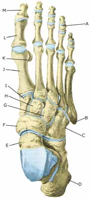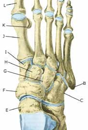
BONE FRACTURE IN THE ANKLE
|
||
|
||
| Cause: A blow or violent twist can cause a fracture of the bone (X-ray picture).
Symptoms: Pain when applying pressure (direct or indirect tenderness), and when applying load or strain. Acute treatment: Click here. Examination: X-ray examination will usually reveal the fracture. The fracture can in some cases first be seen after 14 days, thus the x-ray examination should be repeated if there is a continued suspicion of a fracture. Treatment: Treatment is completely dependent upon which bones are broken, and whether there is a dislocation of the fracture. In some cases relief and rest without bandaging can be opted for, whereas other types of fracture require bandaging and possibly surgical intervention (article) (X-ray picture). Rehabilitation: Rehabilitation is totally dependent upon the type of fracture, and the treatment (conservative or surgical). Complications: If there is not a steady improvement in the condition a medical examination should be performed once more to ensure that the fracture is healing according to plan. In some cases, a false joint can develop which will require (renewed) surgical treatment (X-ray picture). Special: As there is a risk that the injury can cause permanent disability, all cases should be reported to your insurance company. |