|
||
|
||
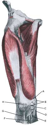
| Cause: Luxation of the knee cap can occur following a blow on the knee, but more often after a sudden and violent knee movement, where the knee is twisted and stretched at the same time. This can cause the knee cap to be displaced to the external side of the knee, whereby the ligaments holding the knee cap will rupture. The knee cap will often impact with the thigh bone, producing the risk of cartilage damage in the knee: cartilage damage in the joint (osteokondrale lesion), cartilage damage on the knee cap (chondromalacia patellae).
Symptoms: Sudden insetting severe pain that renders continued sports activity impossible. The knee cap can become completely displaced to the external side of the knee in some cases, and the knee will consequently be locked in a flexed position (total luxation) until the knee cap suddenly slips into place again allowing the knee to be stretched once more. In other cases, the knee cap will only be partially displaced to the external side of the knee (subluxatio patellae). Acute treatment: Click here. Examination: The diagnosis can be difficult if the knee cap is in its correct position, and anyone with suspicions of displacement of the knee cap should always seek medical attention to ensure the diagnosis and correct treatment. The examination will typically provoke severe pain when the knee cap is pressed outwards (lateral) whilst the knee is flexed (Apprehension test). The knee cap will often be able to be pressed further out on an injured knee than a healthy one. An MR scan will be able to reveal more information regarding the cartilage condition in the knee after a partial or total luxation of the knee cap (article). Treatment: Partial luxation should primarily be treated with relief and rehabilitation. There is no general consensus of opinion regarding treatment of total luxation of the knee cap, some recommend surgery whilst others advocate relief and rehabilitation (article 1) (article 2). Surgery should however be considered with repeated (total or partial) luxation. Rehabilitation of non-operated, partial luxation of the knee cap (subluxatio patellae). Bandage: Tape and bandaging has no documented preventive effect subsequent to previous total or partial luxation of the knee cap (article), however experience has shown that this is utilised to a large degree (tape-instruction). Complications: In case of lack of progress it should be considered whether the diagnosis is correct. Special consideration should be given to:
Special: Since there is a risk that the injury can cause permanent disability, the injury should be reported to your insurance company. A running style analysis can be considered to evaluate whether a correction to the running style can be recommended. |