|
||
|
||
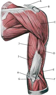
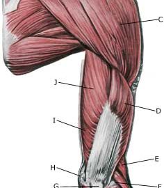
| Cause: When a muscle is suddenly subjected to a load beyond the strength of the muscle, a rupture occurs. The vast majority of triceps ruptures are partial muscle ruptures. Ruptures most often occur if the muscle is contracting while being stretched, e.g. a fall on a bent arm (eccentric contraction).
Symptoms: In light cases a local tenderness is felt after the load (“strained muscle”, “imminent pulled muscle”). In severe cases sudden shooting pains are felt in the muscle (“partial muscle rupture”, “pulled muscle”) and in the worst case a violent snap is felt, after which bending of the elbow against resistance has been severely reduced (“total muscle rupture”). With muscle injuries the following three symptoms are characteristic: pain upon applying pressure, stretching (stretching the elbow) and activation against resistance (bending the elbow). Acute treatment: Click here. Examination: In light cases with only minimal tenderness, medical examination is not necessarily required. In cases of more pronounced pain or difficulty using the arm (stretching the elbow), a medical examination should be carried out to ensure a correct diagnosis and treatment. It can be necessary to supplement the normal clinical examination with an ultrasound scan (article) or an MRI scan. Treatment: Most partial ruptures are treated with relief and rehabilitation. Only in cases with total or near-total ruptures, particularly in young people, is surgery advised (article). Complications: If smooth progress is not achieved, it should be considered whether the diagnosis is correct or whether complications have arisen. Amongst others the following should be considered: complication for muscular bleeding. |