|
||
|
||
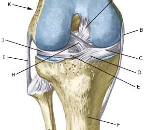
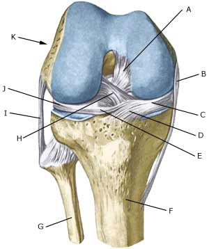
| Cause: Rupture of the posterior cruciate ligament usually occurs following a blow or kick direct on the front of the shin bone just below the knee. (Photo) Symptoms: Usually a snap can be heard or felt and continued sports activity must be aborted. The knee can swell within the first few hours, after which the knee can not bend completely. You can subsequently often sense that the leg gives way (knee failure). Acute treatment: Click here. Examination: If a partial or complete rupture of the cruciate ligament is suspected, you should seek medical attention (casualty ward) immediately, to obtain a diagnosis. The doctor can perform various tests on the knee (rear drawer looseness) to examine the stability of the knee. It should be noted that the looseness in the knee can often only be demonstrated after two weeks. The examination is often more difficult in children and adolescents. It is often necessary to supplement the examination with a MR-scan, ultrasound scan (Ultrasonic image) (article), or arthroscopy to make the diagnosis with certainty. Treatment: Treatment of a rupture of the posterior cruciate ligament usually comprises relief and rehabilitation. It is only in cases of pronounced looseness, or if the rupture is combined with other ligament ruptures, that surgery is recommended (article). A rehabilitation period of at least six months is to be expected. Bandage: Hinge bandages (Don-Joy) can be utilised the first few weeks. Tape treatment of cruciate ligament ruptures in the knee has no sure effect. Complications: If smooth progress is not achieved, it should be considered whether the diagnosis is correct or whether complications have arisen. Amongst others the following should be considered:
Special: Since there is a risk that the injury can cause permanent disability, the injury should be reported to your insurance company. |