Kategoriarkiv: Knee

Inflammation of the kneecap ligament fastening on the kneecap (Sinding-Larsen)

Diagnosis: INFLAMMATION OF THE KNEE CAP LIGAMENT FASTENING ON THE KNEE CAP (SINDING-LARSEN)


Anatomy:
The large anterior thigh muscle (musculus quadriceps femoris) consists of four muscles (M vastus lateralis, M vastus medialis, M vastus intermedius & M rectus femoris). All the muscles fasten on the upper edge of the knee cap. The knee cap ligament (ligamentum patellae) connects the lower edge of the knee cap with the front part of the shin bone (tuberositas tibiae). The function of the knee cap ligament is therefore, when the knee is stretched, to transfer the power the large thigh muscle produces.


  1. M. rectus femoris
  2. M. vastus medialis
  3. Retinaculum patellae mediale
  4. Retinaculum patellae mediale
  5. Tuberositas tibiae
  6. Lig. Patellae
  7. Retinaculum patellae laterale
  8. M. vastus lateralis

KNEE FROM THE FRONT

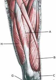
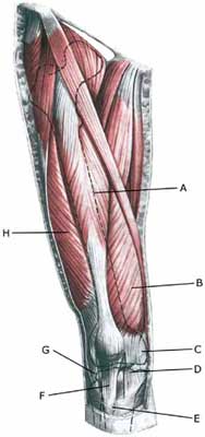
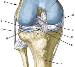
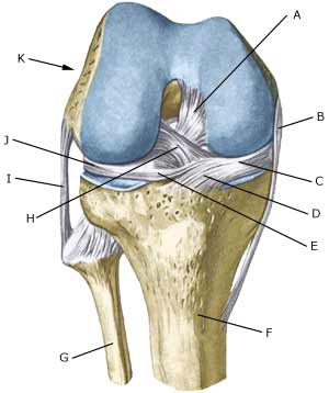
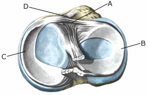
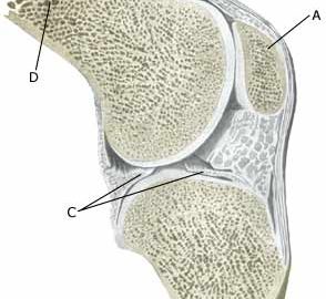
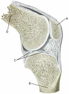
Cause: Repeated uniform loads on the knee cap ligament (jumping, kicking) causes microscopic ruptures at the knee cap ligament fastening on the lower edge of the knee cap. As the load often continues despite tenderness, which in the early stages diminishes after warm-up, a chronic inflammation gradually occurs in the ligament. In some cases, the symptoms will arise at the ligament fastening on the lower edge of the knee cap (Photo)

Symptoms: Jumping and similar activity will initially cause tenderness at the lower edge of the knee cap. The pain diminishes during the first weeks/months after warm-up. If the sport activity continues the pain will increase, resulting in the activity eventually becoming impossible.

Acute treatment: Click here.

Examination: The diagnosis is often made following a normal medical examination, however, if there is any doubt surrounding the diagnosis an ultrasound scan should be performed which will be able to reveal a fraying out (fragmentation) of the bone at the apex of the knee cap (aseptic necrosis of the bone) (article).

Treatment: The treatment consists of relief from the pain inducing activity (jumping, kicking) as soon as possible after the onset of symptoms. The injury can in some cases heal within a few weeks if the treatment is instigated quickly. A rehabilitation period of several months must be anticipated if the pain has been in evidence for some months. Emphasis is put on stretching of the anterior thigh muscle. Ice treatment can be repeated every time tenderness is provoked during the rehabilitation period at the apex of the knee cap.

Rehabilitation of children and adolescents: INSTRUCTION

Bandage: Some patients have the opinion that the application of tape or bandage around the shin bone directly under the knee cap can relieve the discomfort (tape-description).

Complications: In case of lack of progress it should be considered whether the diagnosis is correct or whether complications have arisen. Special consideration should be given to:

Special: Since there is a risk that the injury can cause permanent disability, the injury should be reported to your insurance company.

Osgood-Schlatter desease

Diagnosis: OSGOOD-SCHLATTER DISEASE


Anatomy:
The large anterior thigh muscle (musculus quadriceps femoris) consists of four muscles (m vastus lateralis, m vastus medialis, m vastus intermedius & m rectus femoris). All the muscles fasten on the upper edge of the knee cap. The knee cap ligament (ligamentum patellae) connects the lower edge of the knee cap with the front part of the shin bone (tuberositas tibiae), where children and adolescents have a growth zone. The function of the knee cap ligament is therefore, when the knee is stretched, to transfer the power the large thigh muscle produces.


  1. M. rectus femoris
  2. M. vastus medialis
  3. Retinaculum patellae mediale
  4. Retinaculum patellae mediale
  5. Tuberositas tibiae
  6. Lig. Patellae
  7. Retinaculum patellae laterale
  8. M. vastus lateralis

KNEE FROM THE FRONT


(Photo)

Cause: Repeated uniform loads on the knee cap (jumping, kicking) cause an over-load conditioned inflammation with a fragmentation of the bone at the knee cap ligaments fastening on the growth zone at the upper front part of the shin bone (tuberositas tibiae). The mechanism behind development of Osgood-Schlatter disease is the same in adults as for jumper’s knee. Osgood-Schlatter disease is one of the most common sports injuries in children and adolescents.

Symptoms: Slowly insetting tenderness at the upper, front part of the shin bone (tuberositas tibiae) during and after the sports activity. If the discomfort has a long duration, the bone fastening on the shin bone will become more prominent and can become so large that kneeling will be a problem. It is especially boys in the 10-16 age group that have the symptoms, and the condition is very common and can be seen in almost all boys’ football teams. The symptoms will diminish at around age 17 when the growth zone on the shinbone closes.

Acute treatment: Click here.

Examination: The diagnosis can usually be made with certainty under a normal medical examination, revealing localised tenderness on the knee cap fastening on the upper, front part of the shin bone. If there is any doubt surrounding the diagnosis an ultrasound scan can be performed to identify the changes (ultrasonic image), however, this is seldom necessary in uncomplicated cases (article).

Treatment: The treatment comprises relief from the pain inducing activity (jumping, kicking). The injury can in some cases heal within a few weeks if the treatment is instigated quickly whereas a rehabilitation period of several months must be anticipated if the pain has been in evidence for some months. Emphasis is placed on stretching of the anterior thigh muscle. Ice treatment can be repeated every time tenderness is provoked in the knee cap ligament fastening during the rehabilitation period. If during the rehabilitation period pain is experienced when walking, medicinal treatment in the form of rheumatic medicine (NSAID) in gel form can be considered. Injection of corticosteroid should not be considered in the treatment (article). The sports activity can be cautiously resumed when the pain has diminished. Relapses will often occur, which should be followed as soon as possible with a period of relief. During the relief period it will usually be sufficient to abstain from the most stressful exercises (jumping), whilst many other training exercises can be performed without discomfort. In the majority of cases it will be therefore be possible to participate in at least a part of the sports activity (for example as goal-keeper).

Rehabilitation of children and adolescents: INSTRUCTION

Bandage: Some patients have the opinion that the application of tape or bandage around the shin bone directly under the knee cap can relieve the discomfort (tape-description).

Complications: In case of lack of progress it should be considered whether the diagnosis is correct or whether complications have arisen. Special consideration should be given to:

  • meniscus lesion
  • cartilage damage (osteochondral lesions)
  • bone membrane tear (periosteal avulsion)
  • tendinitis
  • bursitis
  • inflamed mucous fold (plica synovialis)

A tearing of the knee-cap tendon from the fastening on the shinbone has only been described in very rare cases. The torn part of the bone (on which the knee cap tendon is fastened) can be fixed to the shinbone again under surgical operation (article).

Special:
Shock absorbing shoes or insoles will reduce the load on the knee.

Meniscus lesion


Diagnosis: MENISCUS LESION


Anatomy:
The bones in the knee joint comprise the thigh bone (femur), shin bone (tibia) and the knee cap (patella). The articular surfaces on the thigh bone, shin bone and the knee cap are covered with a few mm thick cartilage coating which serves to reduce the strain on the articular surfaces. There is a ring-formed cartilage disk (meniscus) internally and externally in the joint. The knee joint is strengthened by a joint capsule which is in turn strengthened on the sides by an outer and an inner collateral ligament (ligamentum collaterale laterale (LCL) and ligamentum collaterale mediale (MCL)). The internal meniscus is attached to the inner collateral ligament, whereas the external meniscus is not attached to the outer collateral ligament.


  1. Ligamentum cruciatum posterius
  2. Ligamentum collaterale mediale/tibiale
  3. Meniscus medialis
  4. Insertio anterior menisci medialis
  5. Ligamentum transversum genus
  6. Tibiae
  7. Fibulae
  8. Ligamentum cruciatum anterius
  9. Ligamentum collaterale laterale/fibulare
  10. Meniscus lateralis
  11. Femur

KNEE JOINT FROM THE FRONT


  1. Ligamentum transversum genus
  2. Meniscus lateralis
  3. Meniscus medialis
  4. Insertio anterior menisci

KNEE JOINT FROM ABOVE

Cause: Meniscus lesions often occur when the knee is stretched at the same time as rotating. The meniscus will consequently be squeezed between the thigh bone and the shin bone. The inner meniscus is more often damaged than the external. Meniscus injuries in children is very rare (article-1)(article-2)

Symptoms: Pain along the joint line when applying pressure and when rotating the knee. The pain often occurs suddenly when performing certain movements, and can be accompanied by swelling of the knee. In some instances the knee can lock due to a splinter from the meniscus becoming jammed. In other cases, a small and tender outpouching of fluid can be sensed at the joint line (meniscus cyst). Prolonged discomfort will often cause the thigh muscle to diminish.

Acute treatment: Click here.

Examination: A medical examination is always necessary if there is any suspicion of injury within the knee joint. A normal clinical examination is often not sufficient. The doctor can perform various knee tests (meniscus test), however, there is typically tenderness along the joint line which is aggravated when rotating the knee joint. It is often necessary to perform an arthroscopy (telescopic examination of the joint), MR-scan or ultrasound scan to make the diagnosis (article) (ultrasonic image).

Treatment: In uncertain cases the treatment comprises relief and rehabilitation of the knee. If the discomfort does not slowly diminish, if there are instances of “locking” or if it is clear that the meniscus lesion is large (e.g. assessed from an ultrasound scan), surgery should be considered. Under an operation, it can be attempted to sew the torn piece of meniscus back (which will require a considerably longer rehabilitation period, but will probably in the long term reduce the risk of degenerative arthritis in the knee) (article). The torn piece of meniscus is usually removed, but in rare cases the whole meniscus is taken out. Caution is often exercised when recommending removal of the meniscus in children and adolescents, as this can often give prolonged discomfort (article).

Rehabilitation of children and adolescents: INSTRUCTION

Following an operation, exerting load on the knee can commence as soon as the pain and swelling in the knee has diminished. In the best cases, full activity is possible after a period of a few weeks. In uncomplicated cases, it should be possible to resume a full level of sports activity during the course of a month. Rehabilitation must not be allowed to cause increased swelling (or pain) in the knee.

Complications: If insufficient progress is made prior to an operation it must be considered if the diagnosis is correct. Supplementary examinations will often be required (X-ray, ultrasound or MR scan). In particular the following should be considered:

Following an arthroscopy, fluid accumulation in the joint (traumatic arthritis/synovitis) should be considered, and infection in the scar or knee joint, which will always require medical attention as soon as possible.

Special: Since there is a risk that the injury can cause permanent disability, the injury should be reported to your insurance company.


Luxation of the knee cap

Diagnosis: LUXATION OF THE KNEE CAP
(SUB-)LUXATIO PATELLAE)


Anatomy:
The large anterior thigh muscle (musculus quadriceps femoris) consists of four muscles (M vastus lateralis, M vastus medialis, M vastus intermedius & M rectus femoris). All the muscles fasten on the upper edge of the knee cap. The knee cap ligament (ligamentum patellae) connects the lower edge of the knee cap with the front part of the shin bone (tuberositas tibiae). The knee cap is held in place by the structures that fasten on the knee cap, especially the anterior thigh muscle, joint capsule and several ligaments (retiaculum patellae mediale & retiaculum patellae laterale) as well as the knee cap ligament.

  1. Tendo m. adductoris magni
  2. Retinaculum patellae mediale
  3. Meniscus medialis
  4. Ligamentum collaterale mediale/tibiale
  5. Bursa anserina
  6. Bursa subtendinea m. sartori
  7. Ligamentum patellae
  8. Patella

KNEE FROM THE FRONT

Cause: Luxation of the knee cap can occur following a blow on the knee, but more often after a sudden and violent knee movement, where the knee is twisted and stretched at the same time. This can cause the knee cap to be displaced to the external side of the knee, whereby the ligaments holding the knee cap will rupture. The knee cap will often impact with the thigh bone, producing the risk of cartilage damage in the knee: cartilage damage in the joint (osteokondrale lesion), cartilage damage on the knee cap (chondromalacia patellae).

Symptoms: Sudden insetting severe pain that renders continued sports activity impossible. The knee cap can become completely displaced to the external side of the knee in some cases, and the knee will consequently be locked in a flexed position (total luxation) until the knee cap suddenly slips into place again allowing the knee to be stretched once more. In other cases, the knee cap will only be partially displaced to the external side of the knee (subluxatio patellae).

Acute treatment: Click here.

Examination: The diagnosis can be difficult if the knee cap is in its correct position, and anyone with suspicions of displacement of the knee cap should always seek medical attention to ensure the diagnosis and correct treatment. The examination will typically provoke severe pain when the knee cap is pressed outwards (lateral) whilst the knee is flexed (Apprehension test). The knee cap will often be able to be pressed further out on an injured knee than a healthy one. An MR scan will be able to reveal more information regarding the cartilage condition in the knee after a partial or total luxation of the knee cap (article).

Treatment: Partial luxation should primarily be treated with relief and rehabilitation. There is no general consensus of opinion regarding treatment of total luxation of the knee cap, some recommend surgery whilst others advocate relief and rehabilitation (article 1), (article 2). Surgery should however be considered with repeated (total or partial) luxation.

Rehabilitation of children and adolescents: INSTRUCTION

Rehabilitation of non-operated, partial luxation of the knee cap (subluxatio patellae).

Bandage: Tape and bandaging has no documented preventive effect subsequent to previous total or partial luxation of the knee cap (article), however experience has shown that this is utilised to a large degree, (tape-description).

Complications: In case of lack of progress it should be considered whether the diagnosis is correct. Special consideration should be given to:

Inner collateral ligament rupture

Diagnosis: INNER COLLATERAL LIGAMENT RUPTURE
(RUPTURA COLLATERALE LIGAMENTUM MEDIALE/TIBIALE)


Anatomy:
The bones in the knee joint comprise the thigh bone (femur), shin bone (tibia) and the knee cap (patella). There is furthermore a small joint between the shin bone and the calf bone (fibula). The knee joint is strengthened by a joint capsule which is in turn strengthened on the sides by an outer and an inner collateral ligament (ligamentum collaterale laterale (LCL) and ligamentum collaterale mediale (MCL)). Inside the knee there are two ligaments, the anterior and posterior cruciate ligaments (ligamentum cruciatum anterius (ACL) and ligamentum cruciatum posterius (PCL).


  1. Ligamentum cruciatum posterius
  2. Ligamentum collaterale mediale/tibiale
  3. Meniscus medialis
  4. Insertio anterior menisci medialis
  5. Ligamentum transversum genus
  6. Tibiae
  7. Fibulae
  8. Ligamentum cruciatum anterius
  9. Ligamentum collaterale laterale/fibulare
  10. Meniscus lateralis
  11. Femur

KNEE FROM THE FRONT


  1. Tendo m. adductoris magni
  2. Retinaculum patellae mediale
  3. Meniscus medialis
  4. Ligamentum collaterale mediale/tibiale
  5. Bursa anserina
  6. Bursa subtendinea m. sartori
  7. Ligamentum patellae
  8. Patella

KNEE FROM THE FRONT

Cause: When the shin bone is pressed outwards in relation to the thigh bone the inner side-ligament is stretched. If the load becomes sufficiently powerful (as is the case when the load is sudden, so that the thigh muscles do not have time to tighten and stabilize the joint) the ligament can rupture.

Symptoms: Sudden pain on the inside of the knee. Occasionally a snap is felt when the ligament ruptures. In severe cases the athlete complains about a sense of looseness in the knee.

Acute treatment: Click here.

Examination: If a total or partial rupture of the ligaments in the knee is suspected, you should seek medical attention for a diagnosis. An x-ray examination is recommended as many ligament injuries in children and adolescents can be accompanied by damage to the bone at the point of attachment of the ligaments (article). The doctor can perform various knee tests (internal side-instability-test), to examine the stability of the knee. If the knee is stable the injury is called a “sprain” of the inner side-ligament. If the knee is loose the injury is called a “rupture” of the ligament. The diagnosis is usually made from a normal medical examination. If there are any doubts surrounding the diagnosis, an ultrasound scan can be performed which will reveal the rupture and the bleeding along the ligaments (Ultrasonic image) .

Treatment: The treatment of a partial or total rupture of the inner side-ligament involves relief and rehabilitation. If the knee is markedly loose, you can, for a short period, use a support splint (Don Joy). Surgical treatment previously considered has today largely been abandoned in uncomplicated ruptures of the inner side-ligament (article-1)(article-2). (However if the rupture of the inner side-ligament is combined with other ligament ruptures, many would recommend surgery).

Rehabilitation of children and adolescents: INSTRUCTION

Bandage: Hinge bandages can be used initially (Don Joy). Tape treatment of ligament ruptures in the knee have no sure effect.

Complications: If smooth progress is not achieved, it should be considered whether the diagnosis is correct or whether complications have arisen. Supplementary examinations (X-ray, ultrasound, MR scan or arthroscopy) will often be required. The following should be considered:

An inflammation by the ligament fastening is sometimes seen later in the course of the injury, and in some cases bursitis at the ligament fastening is seen (article).

Special: Since there is a risk that the injury can cause permanent disability, the injury should be reported to your insurance company.

Outer collateral ligament rupture

Diagnosis: OUTER COLLATERAL LIGAMENT RUPTURE
(RUPTURA LIGAMENTUM COLLATERALE LATERALE/FIBULARE)


Anatomy:
The bones in the knee joint comprise the thigh bone (femur), shin bone (tibia) and the kneecap (patella). There is furthermore a small joint between the shin bone and the calf bone (fibula). The knee joint is strengthened by a joint capsule which is in turn strengthened on the sides by an outer and an inner collateral ligament (ligamentum collaterale laterale (LCL) and ligamentum collaterale mediale (MCL)). Inside the knee there are two ligaments, the anterior and posterior cruciate ligaments (ligamentum cruciatum anterius (ACL) and ligamentum cruciatum posterius (PCL)).


  1. Ligamentum cruciatum posterius
  2. Ligamentum collaterale mediale/tibiale
  3. Meniscus medialis
  4. Insertio anterior menisci medialis
  5. Ligamentum transversum genus
  6. Tibiae
  7. Fibulae
  8. Ligamentum cruciatum anterius
  9. Ligamentum collaterale laterale/fibulare
  10. Meniscus lateralis
  11. Femur

KNEE FROM THE FRONT


  1. M. gastrocnemius
  2. Caput fibulae
  3. B. subtendinea m. bicipitis femoris inferior
  4. M. biceps femoris
  5. Ligamentum collaterale laterale/fibulare
  6. M. plantaris

OUTER LOWER LEG

Cause: When the shin bone is pressed inwards in relation to the thigh bone the outer side-ligament is stretched. If the load becomes sufficiently powerful (as is the case when the load is sudden, so that the thigh muscles do not have time to tighten and stabilize the joint) the ligament can rupture.

Symptoms: Sudden pain on the outside of the knee. Occasionally a snap is felt when the ligament ruptures. In severe cases the athlete complains about a sense of looseness in the knee

Acute treatment: Click here.

Examination: If a total or partial rupture of the ligaments in the knee is suspected, you should seek medical attention for a diagnosis. An x-ray examination is recommended as many ligament injuries in children and adolescents can be accompanied by damage to the bone at the point of attachment of the ligaments (article). The doctor can perform various knee tests to examine the stability of the knee (external side-instability-test). If the knee is stable the injury is called a “sprain” of the outer side-ligament. If the knee is loose the injury is called a “rupture” of the ligament. The diagnosis is usually made from a normal medical examination. If there are any doubts surrounding the diagnosis, an ultrasoundexamination scan can be performed which will reveal the rupture and the bleeding along the ligaments (Ultrasonic image)

Treatment: The treatment of a partial or total rupture of the outer side-ligament involves relief and rehabilitation (article). If the knee is markedly loose, you can, for a short period, use a support splint (Don Joy). With pronounced sideways looseness surgery can be considered (article). (However if the rupture of the outer side-ligament is combined with other ligament ruptures, as is often the case, surgery is recommended).

Rehabilitation of children and adolescents: INSTRUCTION

Bandage: Hinge bandages can be used initially (Don Joy). Tape treatment of ligament ruptures in the knee have no sure effect.

Complications: If smooth progress is not achieved, it should be considered whether the diagnosis is correct or whether complications have arisen. Supplementary examinations (X-ray, ultrasound, MR scan or arthroscopy) will often be required. The following should be considered:

An inflammation by the ligament fastening is sometimes seen later in the course of the injury, and in some cases bursitis at the ligament fastening is seen (article).

Special: Since there is a risk that the injury can cause permanent disability, the injury should be reported to your insurance company.

Rupture of the anterior cruciate ligament

Diagnosis: RUPTURE OF THE ANTERIOR CRUCIATE LIGAMENT
(RUPTURA LIGAMENTUM CRUCIATUM ANTERIUS)


Anatomy:
The bones in the knee joint comprise the thigh bone (femur), shin bone (tibia) and the knee cap (patella). There is furthermore a small joint between the shin bone and the calf bone (fibula). The knee joint is strengthened by a joint capsule which is in turn strengthened on the sides by an outer and an inner collateral ligament (ligamentum collaterale laterale (LCL) and ligamentum collaterale mediale (MCL)). Inside the knee there are two ligaments, the anterior and posterior cruciate ligaments (ligamentum cruciatum anterius (ACL) and ligamentum cruciatum posterius (PCL)).


  1. Ligamentum cruciatum posterius
  2. Ligamentum collaterale mediale/tibiale
  3. Meniscus medialis
  4. Insertio anterior menisci medialis
  5. Ligamentum transversum genus
  6. Tibiae
  7. Fibulae
  8. Ligamentum cruciatum anterius
  9. Ligamentum collaterale laterale/fibulare
  10. Meniscus lateralis
  11. Femur

KNEE JOINT FROM THE FRONT

Cause: Rupture of the anterior cruciate ligament usually occurs during running with fast changes of direction or as the result of a fall while skiing. Most injuries occur without contact with an opponent. Cruciate ligament injuries in adolescents are being diagnosed with increasingly regularity.

Symptoms: Usually a snap can be heard or felt and continued sports activity must be aborted. The knee swells within the first few hours, after which the knee can not bend completely. You can subsequently often sense that the leg gives way (knee failure).

Acute treatment: Click here.

Examination: If a partial or complete rupture of the cruciate ligament is suspected, you should seek medical attention (casualty ward) immediately, to obtain a diagnosis. The doctor can perform various tests on the knee (front drawer looseness, Lachmann Pivot shift) to examine the stability of the knee. It should be noted that the looseness in the knee can often only be demonstrated after two weeks. The fluid in the knee can be drained. Blood in the knee provides a very strong suspicion of a rupture of the anterior cruciate ligament. The examination is often more difficult in adolescents. The bleeding that occurs after a rupture of the anterior cruciate ligament can usually be seen in an ultrasound scan (Ultrasonic image) (article) or MR-scan. Arthroscopy (a telescopic examination of the knee) is the best suited examination if there is any doubt in the diagnosis.

Treatment: Caution will normally be exercised regarding operating on children in the pre-teenage age group, however, teenagers nearing a fully grown state will usually be advised surgery with the insertion of a new cruciate ligament (article-1) (article-2) (article-3).

Rehabilitation of children and adolescents: INSTRUCTION

An intensive rehabilitation period of at least six months is to be expected. It is important that the knee is stretched completely at least twice a day. The surgeon should be consulted if problems occur with stretching the knee completely

Bandage: Hinge bandages (Don-Joy) can be utilised the first few weeks. Tape treatment of cruciate ligament ruptures in the knee has no sure effect.

Complications: In case of lack of progress you should consider various complications:

In cases where the knee cap tendon is used as a new cruciate ligament an inflammation is often seen where the knee cap tendon has been removed (article).
In children and adolescents there is a risk of affecting the development of the leg operated upon, as it is necessary to pierce the growth zone to insert the artificial cruciate ligament (article).


Special:
Since there is a risk that the injury can cause permanent disability, the injury should be reported to your insurance company.

Rupture of the posterior cruciate ligament

Diagnosis: RUPTURE OF THE POSTERIOR CRUCIATE LIGAMENT
(RUPTURA LIGAMENTUM CRUCIATUM POSTERIUS)


Anatomy:
The bones in the knee joint comprise the thigh bone (femur), shin bone (tibia) and the knee cap (patella). There is furthermore a small joint between the shin bone and the calf bone (fibula). The knee joint is strengthened by a joint capsule which is in turn strengthened on the sides by an outer and an inner collateral ligament (ligamentum collaterale laterale (LCL) and ligamentum collaterale mediale (MCL)). Inside the knee there are two ligaments, the anterior and posterior cruciate ligaments (ligamentum cruciatum anterius (ACL) and ligamentum cruciatum posterius (PCL)).


  1. Ligamentum cruciatum posterius (
  2. Ligamentum collaterale mediale/tibiale
  3. Meniscus medialis
  4. Insertio anterior menisci medialis
  5. Ligamentum transversum genus
  6. Tibiae
  7. Fibulae
  8. Ligamentum cruciatum anterius
  9. Ligamentum collaterale laterale/fibulare
  10. Meniscus lateralis
  11. Femur

KNEE JOINT FROM THE FRONT

Cause: Rupture of the posterior cruciate ligament usually occurs following a blow or kick direct on the front of the shin bone just below the knee.
(Photo)

Symptoms: Usually a snap can be heard or felt and continued sports activity must be aborted. The knee can swell within the first few hours, after which the knee can not bend completely. You can subsequently often sense that the leg gives way (knee failure).

Acute treatment: Click here.

Examination: If a partial or complete rupture of the cruciate ligament is suspected, you should seek medical attention (casualty ward) immediately, to obtain a diagnosis. The doctor can perform various tests on the knee (rear drawer looseness) to examine the stability of the knee. It should be noted that the looseness in the knee can often only be demonstrated after two weeks. The examination is often more difficult in children and adolescents. It is often necessary to supplement the examination with a MR-scan, ultrasound scan (Ultrasonic image) (article), or arthroscopy to make the diagnosis with certainty.

Treatment: Treatment of a rupture of the posterior cruciate ligament usually comprises relief and rehabilitation. It is only in cases of pronounced looseness, or if the rupture is combined with other ligament ruptures, that surgery is recommended (article).

Rehabilitation of children and adolescents: INSTRUCTION

A rehabilitation period of at least six months is to be expected.

Bandage: Hinge bandages (Don-Joy) can be utilised the first few weeks. Tape treatment of cruciate ligament ruptures in the knee has no sure effect.

Complications: If smooth progress is not achieved, it should be considered whether the diagnosis is correct or whether complications have arisen. Amongst others the following should be considered:

Special: Since there is a risk that the injury can cause permanent disability, the injury should be reported to your insurance company.

Cartilage damage in the joint

Diagnosis: CARTILAGE DAMAGE IN THE JOINT
(OSTEOCHONDRALE LÆSIONER, OSTEOCHONDRITIS DISSICANS)


Anatomy:
The bones in the knee joint comprise the thigh bone (femur), shin-bone (tibia) and the knee cap (patella). The articular surfaces of the thigh bone, shin-bone and knee cap are lined with a cartilage covering of a few millimetre’s thickness which serves to reduce the load or strain on the joint surfaces.

Cause: Twists in the knee joint causing the thigh bone and shin bone to collide can cause damage to the cartilage in the knee. In some cases a piece of cartilage can be shed which can wander in the joint (joint-mouse) and become jammed. The synovial membrane can become inflamed, and increase the production of synovial fluid. In other cases, and for unknown reasons, a piece of bone in the knee can loosen (osteochondritis dissicans).

Symptoms: Pain in the joint when under load or strain. Often, periodic swelling of the joint (synovitis).

Examination: Normal medical examination is often not sufficient. To make the diagnosis correctly it is therefore necessary to perform an arthroscopic examination (telescopic examination of the joint), (Photo) or an MR-scan (article-1) (article-2) (article-3). Ultrasound scanning will be able to reveal many, but not all, cartilage damage (Ultrasonic image), which is why an MR-scan is recommended.

Treatment: It is important to make the diagnosis very early if a piece of bone has become loose (osteochondritis dissicans), as the sooner treatment begins, the better the result achieved (article). Treatment can comprise relief or surgery dependant upon the extent of the condition, and whether a piece of bone has possibly become loose (article-1) (article-2). For cartilage damage caused by a twist, the treatment comprises relief from the painful activities until the pain is no longer experienced, after which gradual re-training can be commenced. There is no treatment that can restore the damaged cartilage, which has itself poor restorative ability. Different procedures to enhance the healing can be attempted using arthroscopy, however, the results are generally unsatisfactory. Results from experimental cartilage transplants are still not successful enough to warrant introduction as a routine treatment in the near future. Joint-mouse that provokes the symptoms must be surgically removed. Injection of corticosteroid in the knee joint, performed under ultrasound guidance, can be attempted to combat prolonged and pronounced cases of fluid accumulation in the knee joint (synovitis).

Rehabilitation of children and adolescents: Rehabilitation is completely dependent upon the type of cartilage damage (size and position in the joint) and treatment (conservative or surgical). It is generally attempted to strengthen the musculature around the knee joint.
See: Rehabilitation of children and adolescents in general.

Complications: Large cartilage damage which is positioned on the weight-bearing parts of the joint represents one of the most serious sports injuries, and often results in an end to the sporting career.

Special: As there is a risk that the injury can cause permanent disability, all cases should be reported to your insurance company.

Cartilage damage on the knee cap

Diagnosis: CARTILAGE DAMAGE ON THE KNEE CAP
(CHONDROMALLACIA PATELLAE)


Anatomy:
The bones in the knee joint comprise the thigh bone (femur), shin-bone (tibia) and the knee cap (patella). The articular surfaces of the thigh bone, shin-bone and knee cap are lined with a cartilage covering of a few millimetre’s thickness which serves to reduce the load or strain on the joint surfaces.

  1. Patella
  2. Tibiae
  3. Meniscus lateralis
  4. Femur

KNEE JOINT

Cause: Repeated and uniform loads can cause damage to the cartilage, and subsequently the bone beneath the cartilage (degenerative arthritis). Degenerative arthritis changes (osteoarthrosis) on the rear of the kneecap occur often after a fall on the knee and with many smaller over-loads, however, in many cases the cause is unknown. A weak thigh muscle and increased outward turning of the foot (pronation) have been suspected of being a contributory factor to the disease. The frequency of chondromallacia is dwindling, most probably due to the fact that physicians have become better at making other diagnoses (article).

Symptoms: Pain in the joint when under load or strain, especially with bent knee (for example stairway steps). A sensation of stiffness in the knee after a prolonged period of sitting. Swelling of the joint (synovitis) in occasional cases.

Examination: Normal medical examination is often sufficient. A characteristic of the condition is that the pain occurs when the knee cap presses against the thigh bone, producing a rough, grating sensation. If there are any doubts regarding the diagnosis the examination can be supplemented by X-rays, arthroscopic examination or an MR-scan. (article).

Treatment: Treatment comprises relief from the painful activities until the pain is no longer experienced, after which gradual re-training can be commenced primarily aimed at strengthening the thigh muscles. There is no treatment that can restore the damaged cartilage on the knee cap. For children, treatment of chondromalacia (a softening of the articular cartilage) is non-operative, as the condition has a good prognosis in the younger age groups (article). Several different surgical treatments have been attempted with unsatisfactory results (article).

Rehabilitation of children and adolescents: INSTRUCTION

Bandage: Some patients have felt that the discomfort from cartilage damage behind the knee cap has been helped by applying a knee bandage that holds the knee cap slightly to the side. Alternatively, knee cap stabilising tape can be used, (tape-description).

Complications: If smooth progress is not achieved it should be considered whether the diagnosis is correct, which will often require supplementary examination (X-ray, ultrasound scanning, MR scanning or arthroscopy). The following should especially be considered: