Alle indlæg af urtehave_com

examination-article1

SportNetDoc

Internal impingement in the shoulder of the overhand athlete: pathophysiology, diagnosis, and treatment.

Meister K. Am J Orthop 2000 Jun;29(6):433-8.

The etiology of rotator cuff disease is multifactorial in nature. The process by which the articular surface of the rotator cuff can become diseased secondary to direct abutment against the glenoid rim and labrum has been termed internal impingement. Damage to the undersurface of the rotator cuff can occur from contact at the extremes of shoulder motion and can increase secondary to adaptive changes in bone and soft tissue. Diagnosis is achieved in most instances by a thorough physical examination. Adjunctive tests, particularly magnetic resonance imaging, can increase the accuracy of the diagnosis. If this disease is recognized early, nonoperative intervention may be successful. When nonoperative treatment fails, the use of arthroscopy for the treatment of torn rotator cuff and labral tissue and capsular laxity may be indicated to resolve symptoms and restore the premorbid level of function.

cause-article

SportNetDoc

Internal impingement of the shoulder: a clinical and arthroscopic analysis.

McFarland EG, Hsu CY, Neira C, O’Neil O. J Shoulder Elbow Surg 1999 Sep-Oct;8(5):458-60.

Internal impingement was first described by Walch in 1992 and defined as contact between the supraspinatus tendon and posterior-superior glenoid rim with the shoulder in the cocked, throwing position of 90 degrees of abduction and maximum external rotation. The hypothesis of the study was that this contact may be seen in patients who are not throwing athletes nor in those who have instability. One hundred five consecutive patients who underwent shoulder arthroscopy were prospectively studied with preoperative history and physical examination. All patients underwent general anesthesia and arthroscopy with a standard posterior portal. With the patient under arthroscopy the arm was placed in abduction and external rotation until contact was made or until full elevation was reached. Eighty-five percent (N = 90) of the patients made contact between the rotator cuff and glenoid rim at an average of 95 degrees of abduction and 74 degrees of external rotation. No statistically significant relationship was seen (P > .05) between the position of contact at internal impingement and mechanism of injury, throwing versus nonthrowing, instability, rotator cuff tear, preoperative external rotation, or preoperative impingement signs. The intraoperative finding of contact of the rotator cuff to the posterosuperior glenoid with the arm in abduction and external rotation can occur in a wide spectrum of shoulder disease and is not limited to the throwing athlete. Not all patients with increased laxity and instability demonstrate this contact, suggesting that these factors may not be essential for internal impingement.

treatment-article(slipped-disk)

SportNetDoc

Herniated cervical intervertebralp discs with radiculopathy: an outcome study of conservatively or surgically treated patients.

Heckmann JG, Lang CJ, Zobelein I, Laumer R, Druschky A, Neundorfer B. J Spinal Disord 1999 Oct;12(5):396-401.

To study the functional outcomes of patients with cervical herniated intervertebral discs with radiculopathy but without signs of cervical myelopathy using a retrospective cohort study design. The patients were retrospectively identified by chart reviews. Inclusion criteria were (a) cervical radiculopathy with defined neurological disturbances (sensory disorder, reflex abnormalities, and motor weakness); (b) a spinal computed tomograph scan or magnetic resonance image demonstrating neuroradiologic abnormalities (soft or hard disc); and (c) completion of conservative physical and pharmacological treatment. Of 119 consecutive patients who were treated at the Departments of Neurology and Neurosurgery, University of Erlangen-Nuremberg, between January 1, 1985, and December 31, 1995, 60 patients met the inclusion criteria and were prospectively assessed, with an average follow-up time of 5.5 years. The patients were interviewed with regard to their daily activities, ability to work, and the surgical treatment they had undergone. A complete neurological examination also was performed. In 88.3%, the onset of disease was acute and in 11.7% subacute or chronic. The most common signs and symptoms were brachialgia (98.3%), neck pain (93.3%), sensory disorders (88.3%), reflex abnormalities (61.7%), and motor weakness (51.7%). The neuroimaging procedures demonstrated a disc prolapse (soft disc) in approximately 90% and spondylotic osteophytes (hard disc) in approximately 10%. During an average follow-up time of 5.5 years (range, 4.6 months-10.6 years) 39 (65%) patients had been treated using only conservative methods (COG = conservatively treated group), and 21 (35%) patients had undergone surgery (ventral discectomy) (SUG = surgically treated group). Brachialgia was completely or essentially improved in 100% of the COG and 95.1% of the SUG. Sensory disorders remitted completely or markedly in 97% of the COG and 75% of the SUG. The reflex abnormalities normalized or improved in 59.2% of the COG and in 53.3% of the SUG. Motor weakness improved in 94.1% of the COG and in 50% of the SUG. Neck pain was difficult to treat. It improved in only 36.1% of the COG and in 20% of the SUG. Occupational capacity was lost in 10% of the COG and in 38.9% of the SUG. In a self-rating scale, 89.7% of patients in the COG did not feel disabled in their everyday activities, compared with 66.7% of the patients in the SUG. Patients with a herniated cervical intervertebral disc with radiculopathy can be treated conservatively with good results, although a residual intermittent neck pain syndrome often persists. The patients in the SUG, who initially showed more severe and long-lasting neurological disturbances, were improved at the time of examination, although with more marked residual disorders. Surgery is indicated only when appropriate conservative treatment for a reasonable time has failed.

Slipped disc in the neck

SLIPPED DISC IN THE NAPE OF THE NECK

Diagnosis: SLIPPED DISC IN THE NAPE OF THE NECK
(Prolabsus disci intervertebralis cervicalis)


Anatomy:
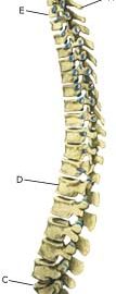
The neck is constructed of box-like vertebrae which are held in place partly with the help of the shape of the bones, and partly by ligaments and muscles. Cartilage discs (disci) are located between the vertebrae which act as shock absorbers. The discs lie close to the spinal canal from where nerves depart to the arms.

  1. Vertebra prominens
  2. Vertebra coccygea I
  3. Promontorium
  4. L I
  5. Th I
  6. Axis

THE BACK SEEN FROM THE SIDE

Cause: If the neck is subjected to a load which exceeds its capabilities, a crack in one of the discs may occur so that the liquid content in the centre of the disc (nucleus pulposus) can be squeezed out and apply pressure on a nerve root.

Symptoms: Pain and stiffness in the neck, radiating to one of the arms. There can be sensory interference and reduction in arm strength. Symptoms are often aggravated by coughing.

Examination: If a slipped disc is suspected medical attention should be sought at once so that special examinations can be performed (extent of sensory interference, reduction of muscle strength, reflexes, foramen compression test) to establish the diagnosis and which treatment should be initiated. In some cases it is necessary to supplement with an MRI scan if the treatment does not progress smoothly.

Treatment: If examination reveals signs of a slipped disc without alarming symptoms (substantial deterioration of muscle strength/paralysis), treatment will primarily be directed at altering the imbalance between the load the neck is subjected to, opposed to the level the neck is trained to manage. It is therefore recommended that you are instructed (possibly by a physiotherapist) in the appropriate way to put a strain on the neck, and which loads and movements should be avoided (“ergotherapeutic guidance”). A few days’ rest and relief may be needed to subdue the pain, after which steadily increasing training should be started with neck and throat muscle exercises. If painkillers are required, paracetamol can be recommended, possibly combined with rheumatic medicine (NSAID). By far the majority of slipped discs can be managed through correct training. In cases where the above treatment does not produce progress in the condition, an MRI scan will be considered with a view to possible operation. Operation is therefore first considered if the rehabilitation programme does not succeed (article), and if the MRI scan can detect a prolapse which can explain the symptoms. In cases with alarming symptoms (substantial deterioration of muscle strength/paralysis) acute hospitalisation is recommended for evaluation of the need for acute surgery.

Special: Training should be performed on a “lifelong” basis to reduce the risk of relapse after a successful rehabilitation. Smoking causes increased risk of pain by reducing the flow of blood to the cartilage discs (disci), implying that daily small injuries do not heal so well. Stopping smoking is therefore an important part of the treatment.

KONDITION

step4

Training ladder for:
NERVE ENTRAPMENT ON THE BACK OF THE SHOULDER BLADE
(ENTRAPMENT N SUPRASCAPULARIS)

STEP 4

KONDITION
Unlimited: Cycling. Swimming. Running.

UDSPÆNDING
(10 min)

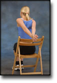
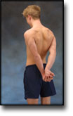
Sit on a chair with your arms outstretched behind your back with hands together. Lift your arms up and backwards so that the front of the shoulders becomes increasingly stretched. Hold the position for 20 seconds and relax for 20 seconds before repeating.

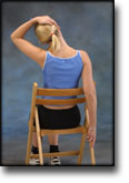
Sit on a chair with one hand on your head and the other holding the chair seat. Slowly draw your head to the side while resisting with the opposite arm so that the muscles on the side of the neck become increasingly stretched. Draw your head in different directions so that all the muscles around the neck are stretched. Hold the position for 20 seconds and relax for 20 seconds before repeating.

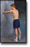
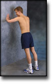
Stand in a doorframe. Press your arms against the frame so that the front of your shoulders become increasingly stretched. Move your arms up and down the doorframe so that different parts of your muscles are stretched. Hold the position for 20 seconds and relax for 20 seconds before repeating.

Stand with your hands together behind your back. Draw your shoulder blades together (imagine trying to hold a pencil between your shoulder blades). Hold the position for 10 seconds and rest for 10 seconds before repeating.

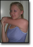
Stand with the injured arm in front of your body. With the opposite hand, press the elbow of the injured arm towards the opposite shoulder, so that the upper part of the arm and the outer shoulder experiences increased stretching. Hold the position for 20 seconds and relax for 20 seconds before repeating.

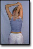
Bend your injured arm behind your head and using the opposite hand, pull the elbow of the injured arm towards the opposite shoulder so that you feel increased stretching. Hold the position for 20 seconds and relax for 20 seconds before repeating.

STYRKE
(50 min)

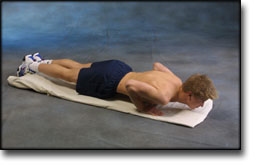
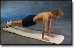
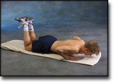
Lie on the floor with instep stretched. Support on your toes and do push-ups without your stomach touching the floor.

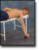
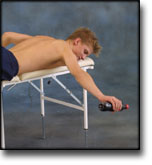
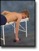
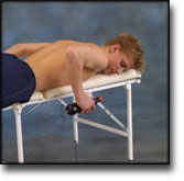
Lie on your stomach on a bench or table with the injured arm hanging over the edge. Raise the injured arm stretched horizontally while drawing the shoulder blade in towards your body. A weight or bottle can be held in the hand to increase the load.

Lie on your stomach on a bench or table with the injured arm hanging over the edge holding a weight or bottle. Slowly draw the hand up to the shoulder joint.

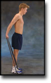
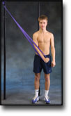
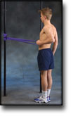
Stand with the elastic under your foot. Hold the elastic with the injured arm and drawn the arm slowly back and upwards so that the elastic is taut.

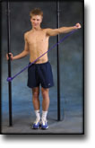
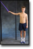
Stand with the good shoulder against a wall. Hold the elastic with the injured arm and move the outstretched arm to the side and away from your body so that the elastic becomes taut.

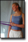
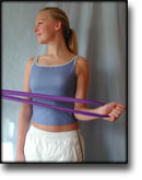
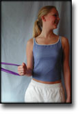
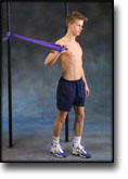
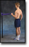
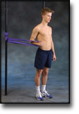
Stand holding the elastic with the upper arm against your body and elbow bent at 90 degrees. Twist your lower arm outwards so that the elastic is taut and draw your arm slowly back again. The elbow must be held against your body the whole time.

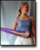
Stand holding the elastic with the upper arm against your body and elbow bent at 90 degrees. Twist your lower arm in over your stomach so that the elastic is taut and draw your arm slowly back again. The elbow must be held against your body the whole time.

Stand with your side against a wall holding the elastic with the injured arm. Stretch the elbow with the upper arm at 90 degrees to your body and the hand above shoulder height. Draw the arm downwards and in over your stomach.

Stretching is carried out in the following way: stretch the muscle group for 3-5 seconds. Relax for 3-5 seconds. The muscle group should subsequently be stretched for 20 seconds. The muscle is allowed to be tender, but must not hurt. Relax for 20 seconds, after which the procedure can be repeated. The time consumed for stretching, coordination and strength training can be altered depending on the training opportunities available and individual requirements.

KONDITION

step3

Training ladder for:
NERVE ENTRAPMENT ON THE BACK OF THE SHOULDER BLADE
(ENTRAPMENT N SUPRASCAPULARIS)

STEP 3

KONDITION
Unlimited: Cycling. Swimming. Running.

UDSPÆNDING
(10 min)

Sit on a chair with your arms outstretched behind your back with hands together. Lift your arms up and backwards so that the front of the shoulders becomes increasingly stretched. Hold the position for 20 seconds and relax for 20 seconds before repeating.

Sit on a chair with one hand on your head and the other holding the chair seat. Slowly draw your head to the side while resisting with the opposite arm so that the muscles on the side of the neck become increasingly stretched. Draw your head in different directions so that all the muscles around the neck are stretched. Hold the position for 20 seconds and relax for 20 seconds before repeating.

Stand in a doorframe. Press your arms against the frame so that the front of your shoulders become increasingly stretched. Move your arms up and down the doorframe so that different parts of your muscles are stretched. Hold the position for 20 seconds and relax for 20 seconds before repeating.

Stand with your hands together behind your back. Draw your shoulder blades together (imagine trying to hold a pencil between your shoulder blades). Hold the position for 10 seconds and rest for 10 seconds before repeating.

Stand with the injured arm in front of your body. With the opposite hand, press the elbow of the injured arm towards the opposite shoulder, so that the upper part of the arm and the outer shoulder experiences increased stretching. Hold the position for 20 seconds and relax for 20 seconds before repeating.

Bend your injured arm behind your head and using the opposite hand, pull the elbow of the injured arm towards the opposite shoulder so that you feel increased stretching. Hold the position for 20 seconds and relax for 20 seconds before repeating.

STYRKE
(50 min)

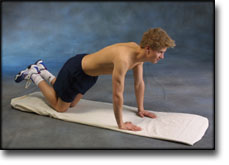
Go down on all fours. Lift your toes from the floor and do push-ups.

Hold an elastic band with the injured arm at a 90-degree angle from the body. Slowly draw the elastic towards yourself so that it tightens.

Hold an elastic band in the good arm. Take hold of the other end of the elastic with the injured arm and draw the injured arm downwards.

Put the elastic under your foot, and with the injured arm draw the other end upwards by bending your arm.

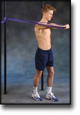
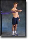
Hold the elastic with the injured arm with elbow bent and your hand at shoulder level. Stretch your arm forwards so that the elastic is tightened.

Stand with your side against a wall. Hold the elastic with the injured arm with elbow bent, upper arm 90 degrees away from your body and your hand at shoulder height. The palm of your hand should face the floor. Drawn your arm downwards and in front of your stomach.

Stand with the elastic under your foot. Hold the elastic with the injured arm and drawn the arm slowly back and upwards so that the elastic is taut.

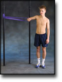
Stand with the good shoulder against a wall. Hold the elastic with the injured arm and move the outstretched arm to the side and away from your body so that the elastic becomes taut.

Stand holding the elastic with the upper arm against your body and elbow bent at 90 degrees. Twist your lower arm outwards so that the elastic is taut and draw your arm slowly back again. The elbow must be held against your body the whole time.

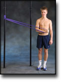
Stand holding the elastic with the upper arm against your body and elbow bent at 90 degrees. Twist your lower arm in over your stomach so that the elastic is taut and draw your arm slowly back again. The elbow must be held against your body the whole time.

Stretching is carried out in the following way: stretch the muscle group for 3-5 seconds. Relax for 3-5 seconds. The muscle group should subsequently be stretched for 20 seconds. The muscle is allowed to be tender, but must not hurt. Relax for 20 seconds, after which the procedure can be repeated. The time consumed for stretching, coordination and strength training can be altered depending on the training opportunities available and individual requirements.

KONDITION

step2

Training ladder for:
NERVE ENTRAPMENT ON THE BACK OF THE SHOULDER BLADE
(ENTRAPMENT N SUPRASCAPULARIS)

STEP 2

KONDITION
Unlimited: Cycling. Swimming. Running.

UDSPÆNDING
(10 min)

Sit on a chair with your arms outstretched behind your back with hands together. Lift your arms up and backwards so that the front of the shoulders becomes increasingly stretched. Hold the position for 20 seconds and relax for 20 seconds before repeating.

Sit on a chair with one hand on your head and the other holding the chair seat. Slowly draw your head to the side while resisting with the opposite arm so that the muscles on the side of the neck become increasingly stretched. Draw your head in different directions so that all the muscles around the neck are stretched. Hold the position for 20 seconds and relax for 20 seconds before repeating.

Stand in a doorframe. Press your arms against the frame so that the front of your shoulders become increasingly stretched. Move your arms up and down the doorframe so that different parts of your muscles are stretched. Hold the position for 20 seconds and relax for 20 seconds before repeating.

Stand with your hands together behind your back. Draw your shoulder blades together (imagine trying to hold a pencil between your shoulder blades). Hold the position for 10 seconds and rest for 10 seconds before repeating.

Stand with the injured arm in front of your body. With the opposite hand, press the elbow of the injured arm towards the opposite shoulder, so that the upper part of the arm and the outer shoulder experiences increased stretching. Hold the position for 20 seconds and relax for 20 seconds before repeating.

Bend your injured arm behind your head and using the opposite hand, pull the elbow of the injured arm towards the opposite shoulder so that you feel increased stretching. Hold the position for 20 seconds and relax for 20 seconds before repeating.

STYRKE
(50 min)

Support with both hands against a wall. Move slowly towards the wall and push away again. The exercise is performed like standing push-ups.

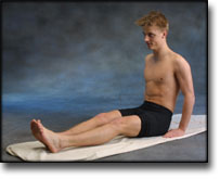
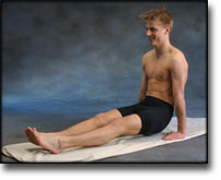
Sit on the floor with outstretched legs. Support with both hands on the floor, and lift yourself by using your arms. Both arms should be stretched.

Hold an elastic band with the injured arm, with the arm by your side and the elbow bent. Slowly draw the elastic towards yourself so that the elastic tightens. The elbow must be bent the whole time.

Hold the elastic with the injured arm with your upper arm alongside your body with the elbow bent. Stretch your arm forwards so that the elastic tightens.

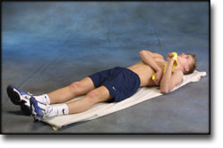
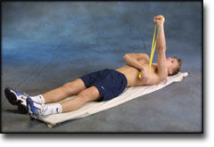
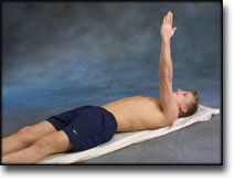
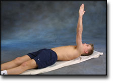
Lie on your back with the injured arm raised upwards. Hold the elastic between your hands with the good arm against your chest. Stretch the injured arm further upwards so that the shoulder blade lifts from the floor.

Stand with the elastic under your foot. Hold the elastic with the injured arm and draw your shoulder upwards. The arm should be kept stretched in against your body the whole time.

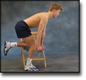
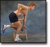
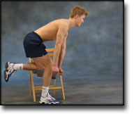
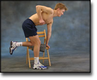
Stand slightly bent over a chair, with your weight on the good arm and the injured arm hanging loosely downwards holding a weight or a filled bottle. Lift your arm/shoulder upwards by using the shoulder blade’s muscles. Using heavier objects or weights can increase the load.

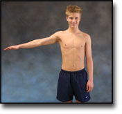
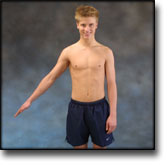
Stand with the injured arm stretched down against your body. Move your arm out from your body to maximum 90 degrees.

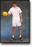
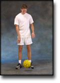
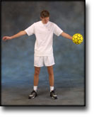
Stand and bounce a ball on the floor backwards and forwards from the injured to the good hand.

Stand holding the elastic with the upper arm against your body and elbow bent at 90 degrees. Twist your lower arm outwards so that the elastic is taut and draw your arm slowly back again. The elbow must be held against your body the whole time.

Stand holding the elastic with the upper arm against your body and elbow bent at 90 degrees. Twist your lower arm in over your stomach so that the elastic is taut and draw your arm slowly back again. The elbow must be held against your body the whole time.

Stretching is carried out in the following way: stretch the muscle group for 3-5 seconds. Relax for 3-5 seconds. The muscle group should subsequently be stretched for 20 seconds. The muscle is allowed to be tender, but must not hurt. Relax for 20 seconds, after which the procedure can be repeated. The time consumed for stretching, coordination and strength training can be altered depending on the training opportunities available and individual requirements.

KONDITION

step1

Training ladder for:
NERVE ENTRAPMENT ON THE BACK OF THE SHOULDER BLADE
(ENTRAPMENT N SUPRASCAPULARIS)

STEP 1

The indications of time after stretching, coordination training and strength training show the division of time for the respective type of training when training for a period of one hour. The time indications are therefore not a definition of the daily training needs, as the daily training is determined on an individual basis.

KONDITION
Unlimited: Cycling. Running.

UDSPÆNDING
(10 min)

Sit on a chair with your arms outstretched behind your back with hands together. Lift your arms up and backwards so that the front of the shoulders becomes increasingly stretched. Hold the position for 20 seconds and relax for 20 seconds before repeating.

Sit on a chair with one hand on your head and the other holding the chair seat. Slowly draw your head to the side while resisting with the opposite arm so that the muscles on the side of the neck become increasingly stretched. Draw your head in different directions so that all the muscles around the neck are stretched. Hold the position for 20 seconds and relax for 20 seconds before repeating.

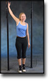
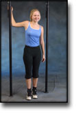
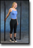
Stand in a doorframe. Press your arms against the frame so that the front of your shoulders become increasingly stretched. Move your arms up and down the doorframe so that different parts of your muscles are stretched. Hold the position for 20 seconds and relax for 20 seconds before repeating.

Stand with your hands together behind your back. Draw your shoulder blades together (imagine trying to hold a pencil between your shoulder blades). Hold the position for 10 seconds and rest for 10 seconds before repeating.

Stand with the injured arm in front of your body. With the opposite hand, press the elbow of the injured arm towards the opposite shoulder, so that the upper part of the arm and the outer shoulder experiences increased stretching. Hold the position for 20 seconds and relax for 20 seconds before repeating.

Bend your injured arm behind your head and using the opposite hand, pull the elbow of the injured arm towards the opposite shoulder so that you feel increased stretching. Hold the position for 20 seconds and relax for 20 seconds before repeating.

STYRKE
(50 min)

Support with both hands against a wall. Put your weight on your hands and draw your shoulder blades backwards and forwards.

Stand slightly bent over a chair, with your weight on the good arm and the injured arm hanging loosely downwards. Lift your arm/shoulder upwards by using the shoulder blade’s muscles. You can bend your arm if you have difficulty in locating the shoulder blade muscles.

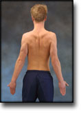
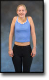
Stand with your arms outstretched at your sides with the palms of your hands facing forwards. Draw your shoulder blades together (imagine trying to hold a pencil between your shoulder blades). Hold the position for 10 seconds and rest for 10 seconds before repeating.

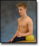
Sit at a table with the injured arm’s elbow on a ball. Press against the ball with a slow movement for 5 seconds. Rest for 5 seconds before repeating.

Lie on your back with the injured arm pointing upwards. Stretch the arm further up so that the shoulder blade lifts from the floor, and go down again. The arm must be outstretched the whole time.

Lift both shoulders slowly upwards and down again.

Stand with the injured arm stretched down against your body. First move the arm out from your body 20 degrees and back again, and then out 40 degrees and back again. Finally move the arm out 60 degrees from your body and back again.

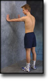
Stand with the injured arm against a wall with elbow bent. Press the arm against the wall and hold the pressure for 10 seconds. Rest for 10 seconds before repeating. Repeat the exercise 10 times.

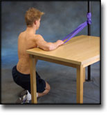
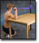
Kneel facing the wall with the injured arm on a table. The tabletop must be at shoulder height. Hold the elastic with the palm facing the wall and move your elbow up and down.

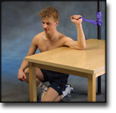
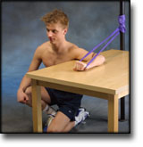
Kneel facing away from the wall with the injured arm on a table. The tabletop must be at shoulder height. Hold the elastic with the back of your hand facing the wall and move your elbow up and down.

Stretching is carried out in the following way: stretch the muscle group for 3-5 seconds. Relax for 3-5 seconds. The muscle group should subsequently be stretched for 20 seconds. The muscle is allowed to be tender, but must not hurt. Relax for 20 seconds, after which the procedure can be repeated. The time consumed for stretching, coordination and strength training can be altered depending on the training opportunities available and individual requirements.

treatment-article2

SportNetDoc

Scapular pain and supra-scapular neuropathy in sports medicine.

Ochsner F, Bonnard C, Chauplannaz G, Kuntzer T. Neurophysiol Clin 2000 Feb;30(1):21-6.

Eight patients with shoulder pain are reported with a history of athletic activities. On examination, performed with a delay of several months, all patients had painful paresis and atrophy of spinati fossa. Electroneuromyography was carried out in all cases and showed a suprascapular nerve axonal loss from the spinati muscles or infraspinatus muscle, signs of denervation-reinnervation in spinati or infraspinatus muscles, normal examination of other scapular girdle muscles, and a coordinate spinati contraction with shoulder displacement excluding rotator cuff tears. All patients had conservative treatment and only two improved. Six patients underwent surgical decompression of the suprascapular nerve; in three, motor function clearly improved, and in three others pain improved. The factors leading to entrapment include stretch mechanisms associated with shoulder movements, leading to suprascapular nerve liability to mechanical lesions. In patients with shoulder pain, the authors recommend an early electrophysiological work-up to recognize an isolated suprascapular neuropathy. The surgical decompression of the nerve should be based on persistent shoulder pain after conservative treatment.