Alle indlæg af urtehave_com

complication-article

SportNetDoc

Acute elbow dislocation: evaluation and management.

Cohen MS, Hastings H 2nd. J Am Acad Orthop Surg 1998 Jan-Feb;6(1):15-23

Most elbow dislocations are stable after closed reduction. Treatment with an early range-of-motion program generally leads to favorable results. Care must be taken to rule out neurovascular involvement and associated osseous or ligamentous injury in the wrist. Late elbow instability and stiffness are rare after simple dislocations. Complex elbow dislocations with associated fractures may require surgical intervention to obtain joint stability; ligament and/or fracture repair is frequently necessary in this situation. Larger periarticular fractures adversely affect functional results. Potential late complications of elbow dislocation include posttraumatic stiffness, posterolateral joint instability, ectopic ossification, and occult distal radioulnar joint disruption.

treatment-article2

SportNetDoc

Return to Australian rules football after acute elbow dislocation:
A report of three cases and review of the literature.

Verrall GM. J Sci Med Sport 2001 Jun;4(2):245-50

Acute elbow dislocation is an uncommon problem encountered in contact sports such as Australian Rules Football [ARF]. Previously there have been few guidelines presented as to when the athlete can safely return to sport following such an injury. During the 1998 playing season the author as medical officer of a professional ARF team encountered three athletes who had an elbow dislocation. All resulted from a fall on an outstretched hand during competition. The athletes returned to football at 13 days, 21 days and 7 days respectively. All subsequently completed the football season without re-injury and at post-season clinical review and one year subsequent to this no athlete described residual symptoms nor was there any loss of range of motion of the elbow joint. Clinical recommendations that allow for athletes to make a safe and early return to contact sport following an episode of acute elbow dislocation include; 1) commencing active mobilization as soon as possible after injury, 2) using passive mobilization to attain full extension as soon as possible, 3) allowing the athlete to return to training before full extension is achieved and 4) allowing the athlete to return to contact sport as soon as full extension is achieved with assistance of elbow stability taping.

treatment-article1

SportNetDoc

Indications for operation in elbow dislocation.

Walter E, Holz U, Kohle H. Orthopade 1988 Jun;17(3):306-12

This paper briefly presents the anatomy of the elbow joint, its pathophysiology, the various types of dislocation of the elbow joint and concomitant injuries, the appropriate diagnostic procedures and the indications for surgery and the operative technique applied for correction of dislocation. The only urgent indications for operative treatment are elbow joint dislocation with concomitant bone injuries, persistent instability or luxation position, open injuries and vessel and/or nerve injuries. For purely ligamentous lesions combined with relative loss of stability an operative procedure does not seem necessary. The operative technique applied for the treatment of habitual or recurrent dislocation is also described.

examination-article

SportNetDoc

Sonography compared with radiography in revealing acute rib fracture.

Griffith JF, Rainer TH, Ching AS, Law KL, Cocks RA, Metreweli C. AJR Am J Roentgenol 1999 Dec;173(6):1603-9.

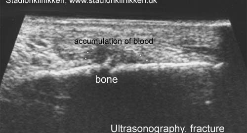
OBJECTIVE: This study was undertaken to compare the sensitivities of sonography and radiography for revealing acute rib fracture. SUBJECTS AND METHODS: Chest radiography and rib sonography were performed on 50 patients with suspected rib fractures. Sonography was performed with a 9- or 12-MHz linear transducer. Fractures were identified by a disruption of the anterior margin of the rib, costochondral junction, or costal cartilage. The incidence, location, and degree of displacement of fractures revealed by radiography and sonography were compared. Sonography was performed again after 3 weeks in 37 subjects. RESULTS: At presentation, radiographs revealed eight rib fractures in six (12%) of 50 patients and sonography revealed 83 rib fractures in 39 (78%) of 50 patients. Seventy-four (89%) of the 83 sonographically detected fractures were located in the rib, four (5%) were located at the costochondral junction, and five (6%) in the costal cartilage. Repeated sonography after 3 weeks showed evidence of healing in all reexamined fractures. Combining sonography at presentation and after 3 weeks, 88% of subjects had sustained a fracture. CONCLUSION: Sonography reveals more fractures than does radiography and will reveal fractures in most patients presenting with suspected rib fracture. Further scientific studies are needed to clarify the appropriate role for sonography in rib fracture detection.

cause-article

SportNetDoc

Stress fractures of the ribs in golfers.

Lord MJ, Ha KI, Song KS. Am J Sports Med 1996 Jan-Feb;24(1):118-22.

During a collaborative review at three institutions, we documented 19 cases of stress fractures of the ribs in golfers. There were 13 men and 6 women with an average age of 39 years (range, 29 to 51). The 4th to 6th ribs were the most commonly injured. All fractures occurred along the posterolateral aspect of the ribs, and nine patients had fractures in more than one rib. Sixteen golfers sustained injury on the leading arm side of the trunk. Eighteen golfers were beginners, and the one experienced golfer had dramatically increased his practice time on the driving range before injury. Plain radiographs were usually diagnostic. However, bone scintigraphy was necessary to reach a diagnosis in three cases. A delay in diagnosis of 6 to 8 months occurred in two cases that were originally misdiagnosed as back strains. Stress fractures of the ribs in golfers may be more common than previously realized and may be incorrectly diagnosed as recalcitrant back strains. Based on the findings of other studies, we think fatigue of the serratus anterior is the mechanism of injury. We recommend strengthening the serratus anterior as rehabilitation after this injury and in a general conditioning program for golfers.

morphine type drugs

Paracetalmol

WEAK MORPHINE TYPE DRUGS

Indication. Analgesic (pain killing tablets) can be used on a greatly limited scale to reduce pain in connection with minor injuries where this is a risk of aggravating the injury with continued activity (i.e. bleeding under the nail and the like). The treatment can naturally also be utilized in many other cases with pain present if the sports activity is discontinued (fracture, lumbago and the like).
Some of the drugs within the “weak morphines” group are on the list of prohibited doping substances!

Mechanism of action. Weak morphines are pain killing tablets with a weak morphine-like effect. The effect is not substantially different from paracetamol. There are several different drugs within the “weak morphines” group, which are all absorbed via the intestines. The effect can be expected after approx. ½-1 hour, with a duration of 3-6 hours.

Side effects. The side effects are of the same character (although weaker) as morphine: nausea, vomiting, drowsiness, constipation and dizziness, and therefore extreme caution should be exercised when driving. The drugs present only a limited risk of dependence and addiction. Overdoses can be life threatening.

Contraindication. Pain killers should never be used to allow an athlete continue a sports activity which can bring about a risk of aggravating the injury. As there are different contraindications with all the drugs in the “weak morphines” group, the attending doctor should acquire a good knowledge of the patient before prescribing drugs from this group.

Dose. Dependent upon which drug is used in the treatment.

Conclusion. Weak morphines are almost never indicated in the treatment of sports injuries, as it is possible to achieve almost the same result with non-prescription drugs with considerably less risk attached.

KONDITION

step4

Training ladder for:
FRACTURE OF THE VERTEBRAL ARCH
(spondylolysis)

STEP 4

Instruction from the physiotherapist in correct back posture (ergonomic guidance) is important.
KONDITION
Unlimited: Cycling. Swimming. Running with increasing distance.

UDSPÆNDING
(15 min)

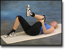
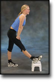
Lie on your back. Draw the injured leg up towards your head so that the muscles in the back of the thigh become increasingly stretched. Perform the exercise with outstretched as well as bent knee. Hold the position for 20 seconds and relax for 20 seconds before repeating. The exercise can also be performed standing with the injured leg outstretched on a chair while the upper body is bent slightly forwards.

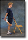
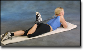
Stand with support from the back of a chair or the wall. Using your hand, bend the knee and draw the foot up and your knee slightly backwards so that the muscles in the front of the thigh become increasingly stretched. Hold the position for 20 seconds and relax for 20 seconds before repeating. The exercise can also be done lying down. If you lie on your stomach you can draw the foot up by using a towel.

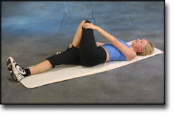
Lie on your back with one leg outstretched and the other bent with the foot on the other side of the outstretched leg. Draw the knee up towards the opposite shoulder so that the buttocks become increasingly stretched. Hold the position for 20 seconds and relax for 20 seconds before repeating.

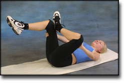
Lie on your back with one leg over the other, and hold behind the lower leg’s knee. Draw the leg up towards your head so that the buttock on the upper leg becomes increasingly stretched. Hold the position for 20 seconds and relax for 20 seconds before repeating.

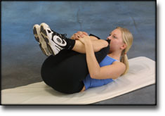
Lie on your back and draw your knee up towards your head while lifting your head so that your back becomes increasingly stretched. Hold the position for 20 seconds and relax for 20 seconds before repeating.

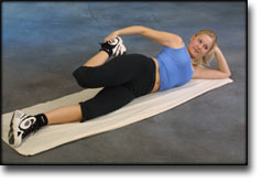
Stand with your hands on your hips and slowly sway your back, pushing slightly with the hands to increase the sway, so that the stomach muscles become increasingly stretched. Hold the position for 20 seconds and relax for 20 seconds before repeating.

KOORDINATION
(5 min)

Seesaw. Balance on two legs, possibly using a hand as support against the wall, balancing subsequently on one leg without support. Look straight ahead and keep knees bent.

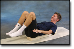
Lie on the floor on a cushion. Bend your knees. Lift your hands from the floor and keep your balance.

STYRKE
(40 min)

Lie on your stomach across a chair and bend both knees. Tighten your buttocks and lift your legs upwards.

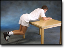
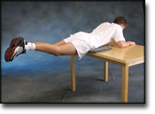
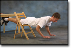
Lie on your stomach across a chair with both feet supported under a tabletop. Lift both hands from the floor and hold the position for 2 seconds. Support with your hands for 2 seconds and repeat the exercise 10 times in quick succession.

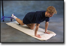
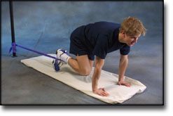
Go down on all fours with elastic around one foot. Attach the elastic to the wall, lift the leg and draw the knee forwards so that the elastic is tightened.

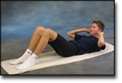
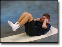
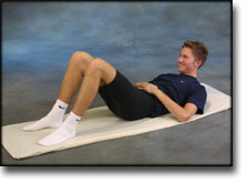
Lie on your back and place your hands behind your neck. Move your head and knees towards each other. Remember not to pull on your neck during the exercise.

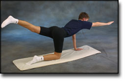
Go down on all fours. Lift right arm and left leg and hold the position for a few seconds, followed by left arm and right leg and hold for a few seconds.

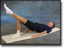
Lie on your back with bent knees. Lift one leg and stretch while at the same time lifting your hip from the floor.

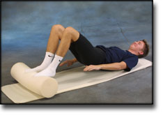
Lie on your back with a ball or firm round cushion under both feet. Roll the ball backwards and forwards in a steady pace while lifting your backside.

Stretching is carried out in the following way: stretch the muscle group for 3-5 seconds. Relax for 3-5 seconds. The muscle group should subsequently be stretched for 20 seconds. The muscle is allowed to be tender, but must not hurt. Relax for 20 seconds, after which the procedure can be repeated. The time consumed for stretching, coordination and strength training can be altered depending on the training opportunities available and individual requirements.

KONDITION

step3

Training ladder for:
FRACTURE OF THE VERTEBRAL ARCH
(spondylolysis)

STEP 3

Instruction from the physiotherapist in correct back posture (ergonomic guidance) is important.
KONDITION
Unlimited: Cycling. Swimming. Running over short distances on a soft surface.

UDSPÆNDING
(15 min)

Lie on your back. Draw the injured leg up towards your head so that the muscles in the back of the thigh become increasingly stretched. Perform the exercise with outstretched as well as bent knee. Hold the position for 20 seconds and relax for 20 seconds before repeating. The exercise can also be performed standing with the injured leg outstretched on a chair while the upper body is bent slightly forwards.

Stand with support from the back of a chair or the wall. Using your hand, bend the knee and draw the foot up and your knee slightly backwards so that the muscles in the front of the thigh become increasingly stretched. Hold the position for 20 seconds and relax for 20 seconds before repeating.

Lie on your back with one leg outstretched and the other bent with the foot on the other side of the outstretched leg. Draw the knee up towards the opposite shoulder so that the buttocks become increasingly stretched. Hold the position for 20 seconds and relax for 20 seconds before repeating.

Lie on your back with one leg over the other, and hold behind the lower leg’s knee. Draw the leg up towards your head so that the buttock on the upper leg becomes increasingly stretched. Hold the position for 20 seconds and relax for 20 seconds before repeating.

Lie on your back and draw your knee up towards your head while lifting your head so that your back becomes increasingly stretched. Hold the position for 20 seconds and relax for 20 seconds before repeating.

Stand with your hands on your hips and slowly sway your back, pushing slightly with the hands to increase the sway, so that the stomach muscles become increasingly stretched. Hold the position for 20 seconds and relax for 20 seconds before repeating.

KOORDINATION
(5 min)

Seesaw. Balance on two legs, possibly using a hand as support against the wall, balancing subsequently on one leg without support. Look straight ahead and keep knees bent.

Lie on the floor on a cushion. Bend your knees. Lift your hands from the floor and keep your balance.

STYRKE
(40 min)

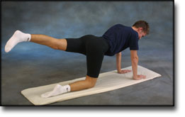
Go down on all fours. Alternately lift and stretch the right and left legs, fully stretching the knee.

Lie on your stomach across a chair and bend both knees. Tighten your buttocks and lift your legs upwards.

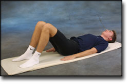
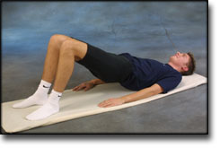
Lie on your back with bent legs. Lift your hip from the floor and hold the position for 5 seconds. Rest for 5 seconds before repeating.

Go down on all fours with elastic around one foot. Attach the elastic to the wall, lift the leg and draw the knee forwards so that the elastic is tightened.

Lie on your back with bent knees. Lift one leg and stretch while at the same time lifting your hip from the floor.

Lie on your back and place your hands behind your neck. Move your head and knees towards each other. Remember not to pull on your neck during the exercise.

Stretching is carried out in the following way: stretch the muscle group for 3-5 seconds. Relax for 3-5 seconds. The muscle group should subsequently be stretched for 20 seconds. The muscle is allowed to be tender, but must not hurt. Relax for 20 seconds, after which the procedure can be repeated. The time consumed for stretching, coordination and strength training can be altered depending on the training opportunities available and individual requirements.

KONDITION

step2

Training ladder for:
FRACTURE OF THE VERTEBRAL ARCH
(spondylolysis)

STEP 2

Instruction from the physiotherapist in correct back posture (ergonomic guidance) is important.
KONDITION
Unlimited: Swimming. Light jogging on a soft surface.

UDSPÆNDING
(15 min)

Lie on your back. Draw the injured leg up towards your head so that the muscles in the back of the thigh become increasingly stretched. Perform the exercise with outstretched as well as bent knee. Hold the position for 20 seconds and relax for 20 seconds before repeating. The exercise can also be performed standing with the injured leg outstretched on a chair while the upper body is bent slightly forwards.

Stand with support from the back of a chair or the wall. Using your hand, bend the knee and draw the foot up and your knee slightly backwards so that the muscles in the front of the thigh become increasingly stretched. Hold the position for 20 seconds and relax for 20 seconds before repeating.

Lie on your back with one leg outstretched and the other bent with the foot on the other side of the outstretched leg. Draw the knee up towards the opposite shoulder so that the buttocks become increasingly stretched. Hold the position for 20 seconds and relax for 20 seconds before repeating.

Lie on your back with one leg over the other, and hold behind the lower leg’s knee. Draw the leg up towards your head so that the buttock on the upper leg becomes increasingly stretched. Hold the position for 20 seconds and relax for 20 seconds before repeating.

Lie on your back and draw your knee up towards your head while lifting your head so that your back becomes increasingly stretched. Hold the position for 20 seconds and relax for 20 seconds before repeating.

Stand with your hands on your hips and slowly sway your back, pushing slightly with the hands to increase the sway, so that the stomach muscles become increasingly stretched. Hold the position for 20 seconds and relax for 20 seconds before repeating.

KOORDINATION
(5 min)

Seesaw. Balance on two legs, possibly using a hand as support against the wall, balancing subsequently on one leg without support. Look straight ahead and keep knees bent.

Lie on the floor on a cushion. Bend your knees. Lift your hands from the floor and keep your balance.

STYRKE
(40 min)

Go down on all fours. Alternately lift and stretch the right and left legs, fully stretching the knee.

Lie on your stomach across a chair with both feet supported under a tabletop. Lift both hands from the floor and hold the position for 2 seconds. Support with your hands for 2 seconds and repeat the exercise 10 times in quick succession.

Lie on your stomach across a chair and bend both knees. Tighten your buttocks and lift your legs upwards.

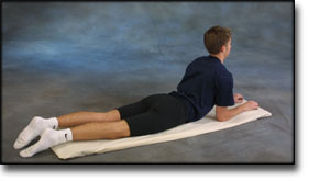
Lie on your stomach. Rise up and support yourself on your elbows keeping your hip against the floor. Hold the position for 20 seconds. Rest for 20 seconds before repeating.

Go down on all fours with elastic around one foot. Attach the elastic to the wall, lift the leg and draw the knee forwards so that the elastic is tightened.

Lie on your back with your hands on your chest. Bend your knees and lift your upper body up from the floor while keeping the small of your back against the floor the whole time.

Stretching is carried out in the following way: stretch the muscle group for 3-5 seconds. Relax for 3-5 seconds. The muscle group should subsequently be stretched for 20 seconds. The muscle is allowed to be tender, but must not hurt. Relax for 20 seconds, after which the procedure can be repeated. The time consumed for stretching, coordination and strength training can be altered depending on the training opportunities available and individual requirements.