Alle indlæg af urtehave_com

Scheuermann disease

Diagnosis: SCHEUERMANN’S DISEASE


Anatomy:
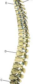
The back is constructed of box-like vertebrae which are held in place partly with the help of the shape of the bones, partly by ligaments and partly by the large and small back and stomach muscles.


  1. Vertebra prominens
  2. Vertebra coccygea I
  3. Promontorium
  4. L I
  5. Th I
  6. Axis

THE BACK SEEN FROM THE SIDE

Cause: Scheuermann’s disease occurring in approximately 4% of the population (article). A curvature of the back occurs (bending over forwards) due to the vertebrae becoming wedge shaped. There are also characteristic x-ray finds. The cause of the condition is unknown, but evidence tends to suggest that the condition is hereditary (article).

Symptoms: Back curvature localised high in the back (thoracal Scheuermann) gives often only few, if any, symptoms. Back curvature localised in the lower back (thoracolumbal or lumbal Scheuermann) does entail back pain for the majority (article).

Examination: The diagnosis is usually made following a medical examination supplemented with an x-ray (at least 3 adjacent vertebrae with at least 5 degrees wedge form, Schmorlske impressions, flattening of discs, irregular end plates) (article). The crooked back is often mistaken in the beginning for “bad posture”. In some cases, CT or MRI scanning is recommended.

Treatment: The vast majority of cases can be treated with training, attempting to maintain the mobility of the back, counteract the curvature tendency and strengthen the stomach and back muscles. A corset can in some cases be used until the young person is fully grown. An operation can be performed only in very rare cases. The condition has a good prognosis (article), and even after an operation it is still possible to take part in many different forms of sport (article).

Rehabilitation of children and adolescents: INSTRUCTION

Complications: In some cases a crooked back can have other causes (infection, nerve disease, inborn bone change, rheumatic illness, bone disease, metabolic disorder).

bandage-article2

SportNetDoc

The incidence of sports-related facial trauma in children.

Perkins SW, Dayan SH, Sklarew EC, Hamilton M, Bussell GS. Ear Nose Throat J 2000 Aug;79(8):632-4, 636, 638.

We conducted a survey of physician members of the American Academy of Facial Plastic and Reconstructive Surgery to determine the incidence and nature of facial traumas seen in their practices. We solicited information on the anatomic location of each injury, the severity of the trauma, and whether the injury occurred during a sports activity. According to the responses, 21% of facial fractures and 29% of nasal fractures were experienced by patients aged 17 years and younger who were participating in sports. We believe that many such injuries can be prevented with greater use of protective equipment.

bandage-article1

SportNetDoc

Management of simple midfacial fractures, particularly in professional soccer players.

Eufinger H, Heise M, Rarreck T. Sportverletz Sportschaden 2000 Mar;14(1):35-40.

Uncomplicated midfacial fractures represent a frequent and typical injury of soccer-players in oral and maxillofacial surgery. The treatment of these fractures in professional players requires special treatment modalities, especially concerning quick rehabilitation. The examples of a nasal bone fracture, a malar bone fracture and a zygomatic arch fracture in 3 professional soccer players demonstrate the therapeutic principles of these most common uncomplicated midfacial fractures. In this context the fabrication of individual facial masks is presented, which allow an improved postoperative protection and therefore an earlier participation of the player in training and matches.

Special(a2)

SportNetDoc

Cerebral concussion in sport. Management and prevention.

Cantu RC. Sports Med 1992 Jul;14(1):64-74.

This article explains the various stresses (tensile, compressive, and shearing) that can affect the brain, and how they may produce the different types of brain injury. The biomechanical forces and dynamics that produce coup versus contra coup injury are covered, as are the common intracranial athletic head injuries, i.e. concussion and the various intracranial haematomas (epidural, subdural, subarachnoid and intracerebral). Though less common in occurrence, because their outcome is so catastrophic, space is also devoted to the recognition, the treatment and (especially in the latter case) the prevention of the malignant brain oedema syndrome of the adolescent and the second impact syndrome of the adult. A major emphasis of this paper is the recognition of the 3 grades of cerebral concussion and the delineation of clear guidelines as to when it is safe to return to collision sports after sustaining such injuries, for the first, second or third time during a given season. Clear guidelines are also presented as to when to discontinue collision sport competition for the remainder of the season after multiple concussions. Because of the concern for the second impact syndrome, the requirement to never allow an athlete with postconcussion syndrome symptoms to return to competition is emphasised. Also covered is the prevention of head injuries, which sports are at greatest risk, and the need for additional research on the cumulative effects of concussion.

Special(a1)

SportNetDoc

Concussion in sports. Guidelines for the prevention of catastrophic outcome.

Kelly JP, Nichols JS, Filley CM, Lillehei KO, Rubinstein D, Kleinschmidt-DeMasters BK. JAMA 1991 Nov 27;266(20):2867-9.

Concussion (defined as a traumatically induced alteration in mental status, not necessarily with loss of consciousness) is a common form of sports-related injury too often dismissed as trivial by physicians, athletic trainers, coaches, sports reporters, and athletes themselves. While head injuries can occur in virtually any form of athletic activity, they occur most frequently in contact sports, such as football, boxing, and martial arts competition, or from high-velocity collisions or falls in basketball, soccer, and ice hockey. The pathophysiology of concussion is less well understood than that of severe head injury, and it has received less attention as a result. We describe a high school football player who died of diffuse brain swelling after repeated concussions without loss of consciousness. Guidelines have been developed to reduce the risk of such serious catastrophic outcomes after concussion in sports.

KONDITION

step4

Training ladder for:
TENDINITIS AT THE ISCHIATIC BONE
(ENTESOPATIA TUBER ISCHIADICUM)

STEP 4

KONDITION
Unlimited: Cycling. Swimming. Running with intermittent sprints.

UDSPÆNDING
(15 min)

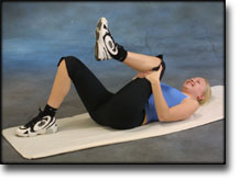
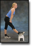
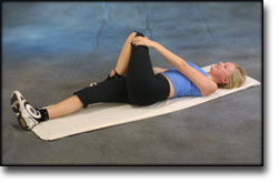
Lie on your back. Draw the injured leg up towards your head so that the muscles in the back of the thigh become increasingly stretched. Perform the exercise with outstretched as well as bent knee. Hold the position for 20 seconds and relax for 20 seconds before repeating. The exercise can also be performed standing with the injured leg outstretched on a chair while the upper body is bent slightly forwards.

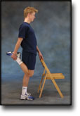
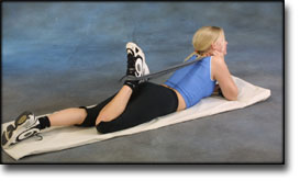
Stand with support from the back of a chair or the wall. Using your hand, bend the knee and draw the foot up and your knee slightly backwards so that the muscles in the front of the thigh become increasingly stretched. Hold the position for 20 seconds and relax for 20 seconds before repeating. The exercise can also be done lying down. If you lie on your stomach you can draw the foot up by using a towel.

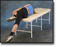
Lie on your side on a table. Bend one leg up under your body and let the other hang over the edge of the table so that the muscles in the outer side of the thigh become increasingly stretched. Hold the position for 20 seconds and relax for 20 seconds before repeating. The exercise can also be done standing by placing the outstretched injured leg behind the good leg at the same time as bending over the injured leg.

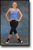
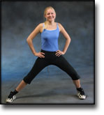
Stand with one leg outstretched and the other slightly bent. Thrust your weight to the side over the bent leg so that the inner side of the opposite thigh becomes increasingly stretched. Hold the position for 20 seconds and relax for 20 seconds before repeating.

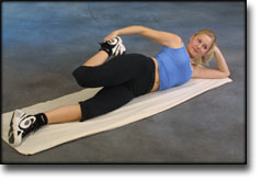
Lie on your back with one leg outstretched and the other bent with the foot on the other side of the outstretched leg. Draw the knee up towards the opposite shoulder so that the buttocks become increasingly stretched. Hold the position for 20 seconds and relax for 20 seconds before repeating.

KOORDINATION
(5 min)

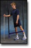
Seesaw. Balance on two legs, possibly using a hand as support against the wall, balancing subsequently on one leg without support. Look straight ahead and keep knees bent.

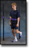
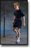
Stand on the healthy leg with elastic fixed around the hip. The elastic should be fixed to the wall or a wall bar. Take-off on the healthy leg and land on the leg to be trained and keep your balance. Remember that the elastic should be positioned so that it gives resistance at the moment of take-off. Change legs.

STYRKE
(40 min)

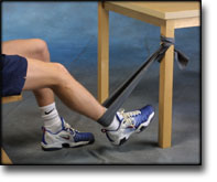
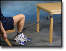
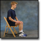
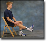
Sit on a chair with elastic around the ankle, facing the elastic. Lift the leg and slowly bend and stretch the knee.

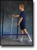
Stand on the healthy leg with the elastic around the inside of the injured leg. Move the injured leg from side to side in a slow smooth movement. Moving the position of the elastic lower down the leg can increase the load.

Stand on the healthy leg with the elastic around the outside of the injured leg. Move the injured leg from side to side in a slow smooth movement. Moving the position of the elastic lower down the leg can increase the load.

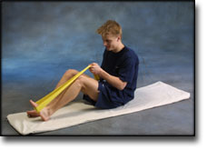
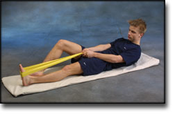
Lie on the floor with slightly bent knees. Put the elastic under the foot and hold firmly with your hands. Stretch the knee so that the elastic is drawn tight and slowly bend the knee again.

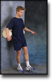
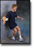
Stand with your back against a wall with a ball or firm round cushion between the wall and your back. Slowly go down to bend your knee 90 degrees before slowly rising up again.

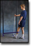
Stand with the elastic around the injured leg, facing towards the elastic. Move the leg backwards and slowly forwards. The elastic can be moved up and down the leg depending upon the strength of the knee – the stronger the knee, the lower the elastic should be.

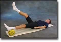
Lie on your back with a ball or firm round cushion under the injured leg. Lift your backside up from the floor and stretch the healthy leg. Hold the position for a few seconds.

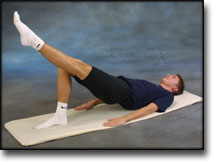
Lie on your back with bent knees. Lift one leg and stretch while at the same time lifting your hip from the floor.

Stretching is carried out in the following way: stretch the muscle group for 3-5 seconds. Relax for 3-5 seconds. The muscle group should subsequently be stretched for 20 seconds. The muscle is allowed to be tender, but must not hurt. Relax for 20 seconds, after which the procedure can be repeated. The time consumed for stretching, coordination and strength training can be altered depending on the training opportunities available and individual requirements.

Treatment(a1)

SportNetDoc

Assessment and management of concussion in sports.

Harmon KG. Am Fam Physician 1999 Sep 1;60(3):887-92, 894.

The most common head injury in sports is concussion. Athletes who sustain a prolonged loss of consciousness should be transported immediately to a hospital for further evaluation. Assessment of less severe injuries should include a thorough neurologic examination. The duration of symptoms and the presence or absence of post-traumatic amnesia and loss of consciousness should be noted. To avoid premature return to play, a good understanding of the possible hazards is important. Potential hazards of premature return to play include the possibility of death from second-impact syndrome, permanent neurologic impairment from cumulative trauma, and the postconcussion syndrome.

Examination(a1)

SportNetDoc

Neuropsychological impairment in amateur soccer players.

Matser EJ, Kessels AG, Lezak MD, Jordan BD, Troost J. JAMA 1999 Sep 8;282(10):971-3.

CONTEXT.
Soccer players incur concussions during matches and training sessions, as well as numerous subconcussive blows to the head from impacts with the soccer ball (headers). The combination of soccer-related concussions and the number of headers may be a risk for chronic traumatic brain injury (CTBI).

OBJECTIVE.
To determine whether amateur soccer players have evidence of CTBI. DESIGN, SETTING, AND PARTICIPANTS: Cross-sectional study of 33 amateur soccer players and 27 amateur athletes involved in swimming and track (controls) in the Netherlands who underwent interviews and neuropsychological testing.

MAIN OUTCOME MEASURES.
Performance of soccer players vs controls on 16 neuropsychological tests having 27 outcomes.

RESULTS.
Compared with control athletes, amateur soccer players exhibited impaired performance on tests of planning (39% vs 13%; P=.001) and memory (27% vs 7%; P=.004). Among soccer players, 9 (27%) had incurred 1 soccer-related concussion and 7 (23%) had had 2 to 5 concussions during their career. The number of concussions incurred in soccer was inversely related to the neuropsychological performance on 6 of the neuropsychological tests.

CONCLUSIONS.
Our results indicate that participation in amateur soccer in general and concussion specifically is associated with impaired performance in memory and planning functions. Due to the worldwide popularity of soccer, these observations may have important public health implications.

Symptoms(a1)

SportNetDoc

Fatalities from head and cervical spine injuries occurring in tackle football: 50 years’ experience.

Mueller FO. Clin Sports Med 1998 Jan;17(1):169-82.

Football head and cervical spine fatalities have been related to 84.9% of all football fatalities from 1945 through 1994. The decade from 1965 through 1974 was responsible for the greatest number and percentage of head and cervical spine fatalities, and the two decades from 1975 through 1994 were associated with the smallest number and percentage. The data reveal that most head and cervical spine fatalities are related to high school football players either tackling or being tackled in a game. Most head fatalities are subdural hematomas, and almost all of the cervical spine fatalities are fractures, dislocations, or fracture-dislocations. There has been a dramatic reduction in these types of fatalities during the last two decades, 1975 through 1994, and the preventive measures that have received most of the credit have been the 1976 rule change that prohibits initial contact with the head and face when blocking and tackling, the NOCSAE helmet standard that went into effect in colleges in 1978 and high schools in 1980, better coaching in the techniques of blocking and tackling, and improved medical care. There has been a reduction of head and cervical spine fatalities, but the analysis of data for the next decade, 1995 through 2004, will reveal the continued effects of the preventive measures discussed and continued research efforts. A number of researchers have stated that in order for the head and cervical spine fatalities to continue decreasing, there must be increased helmet research with an emphasis on concussions and neck injuries. Will the number of fatalities continue to decrease? Only time will tell.

KONDITION

step3

Training ladder for:
TENDINITIS AT THE ISCHIATIC BONE
(ENTESOPATIA TUBER ISCHIADICUM)

STEP 3

KONDITION
Unlimited: Cycling. Swimming. Running with increasing speed.

UDSPÆNDING
(15 min)

Lie on your back. Draw the injured leg up towards your head so that the muscles in the back of the thigh become increasingly stretched. Perform the exercise with outstretched as well as bent knee. Hold the position for 20 seconds and relax for 20 seconds before repeating. The exercise can also be performed standing with the injured leg outstretched on a chair while the upper body is bent slightly forwards.

Stand with support from the back of a chair or the wall. Using your hand, bend the knee and draw the foot up and your knee slightly backwards so that the muscles in the front of the thigh become increasingly stretched. Hold the position for 20 seconds and relax for 20 seconds before repeating. The exercise can also be done lying down. If you lie on your stomach you can draw the foot up by using a towel.

Lie on your side on a table. Bend one leg up under your body and let the other hang over the edge of the table so that the muscles in the outer side of the thigh become increasingly stretched. Hold the position for 20 seconds and relax for 20 seconds before repeating. The exercise can also be done standing by placing the outstretched injured leg behind the good leg at the same time as bending over the injured leg.

Stand with one leg outstretched and the other slightly bent. Thrust your weight to the side over the bent leg so that the inner side of the opposite thigh becomes increasingly stretched. Hold the position for 20 seconds and relax for 20 seconds before repeating.

Lie on your back with one leg outstretched and the other bent with the foot on the other side of the outstretched leg. Draw the knee up towards the opposite shoulder so that the buttocks become increasingly stretched. Hold the position for 20 seconds and relax for 20 seconds before repeating.

KOORDINATION
(5 min)

Seesaw. Balance on two legs, possibly using a hand as support against the wall, balancing subsequently on one leg without support. Look straight ahead and keep knees bent.

Stand on the healthy leg with elastic fixed around the hip. The elastic should be fixed to the wall or a wall bar. Take-off on the healthy leg and land on the leg to be trained and keep your balance. Remember that the elastic should be positioned so that it gives resistance at the moment of take-off. Change legs.

STYRKE
(40 min)

Sit on a chair with elastic around the ankle, facing the elastic. Lift the leg and slowly bend and stretch the knee.

Sit on a chair with elastic attached to the ankle. Raise the leg and slowly stretch and bend the knee.

Stand with your back against a wall with a ball or firm round cushion between the wall and your back. Slowly go down to bend your knee 90 degrees before slowly rising up again.

Stand on the healthy leg with the elastic around the inside of the injured leg. Move the injured leg from side to side in a slow smooth movement. Moving the position of the elastic lower down the leg can increase the load.

Stand on the healthy leg with the elastic around the outside of the injured leg. Move the injured leg from side to side in a slow smooth movement. Moving the position of the elastic lower down the leg can increase the load.

Lie on your back with a ball or firm round cushion under the injured leg. Lift your backside up from the floor and stretch the healthy leg. Hold the position for a few seconds.

Lie on your back with bent knees. Lift one leg and stretch while at the same time lifting your hip from the floor.

Stand with the elastic around the injured leg, facing towards the elastic. Move the leg backwards and slowly forwards. The elastic can be moved up and down the leg depending upon the strength of the knee – the stronger the knee, the lower the elastic should be.

Stretching is carried out in the following way: stretch the muscle group for 3-5 seconds. Relax for 3-5 seconds. The muscle group should subsequently be stretched for 20 seconds. The muscle is allowed to be tender, but must not hurt. Relax for 20 seconds, after which the procedure can be repeated. The time consumed for stretching, coordination and strength training can be altered depending on the training opportunities available and individual requirements.